
Contents
- What You Will Learn
- Key Terms at a Glance
- Tissues: Introduction and Importance
- Levels of Organisation in Living Organisms
- Why Are Plant and Animal Tissues Different?
- Plant Tissues
- Meristematic Tissues
- Permanent Tissues
- Simple and Complex Tissues
- Protective Tissues: Epidermis, Stomata and Cork
- The Three Tissue Systems of a Plant
- Totipotency: When a Permanent Cell Divides Again
- Animal Tissues
- Epithelial Tissue
- Connective Tissue
- Muscular Tissue
- Nervous Tissue
- Animal Tissues at a Glance
- Musculoskeletal System
- Elementary Idea and Joints
- How Tissues Work Together in Movement
- Care: Injuries, Postural Care, Nutrition and Exercise
- Related References
- Yoga and Physical Agility
- Indian Contributions
- Practical: Identifying Tissues from Prepared Slides (Experiment 5)
- Conclusion
- Quick Summary
- Previous Year and Practice Questions
- Frequently Asked Questions
- Continue Reading
- External References
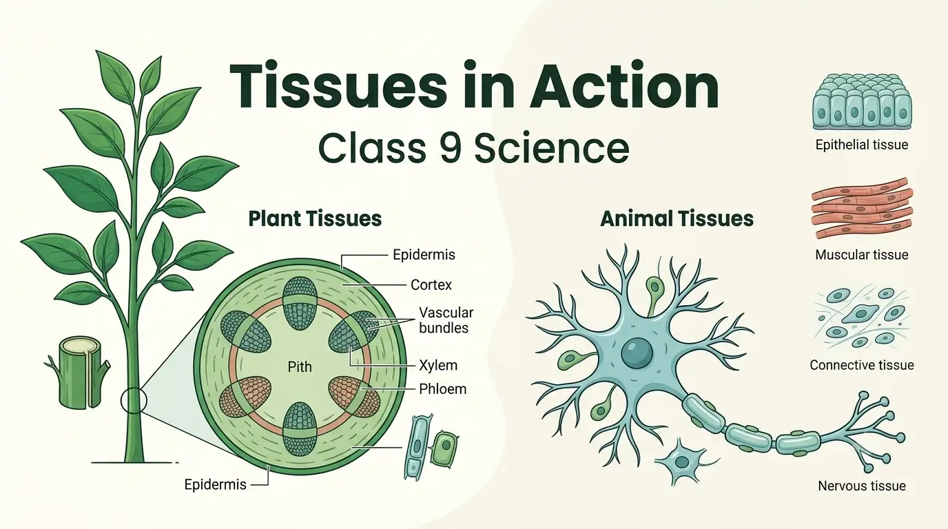
Quick answer: A tissue is a group of cells, similar in structure, that work together to perform a specific function. In plants, the two main groups are meristematic tissues (which keep dividing and make the plant grow) and permanent tissues (simple, complex and protective). In animals, there are four basic tissues: epithelial, connective, muscular and nervous. These notes cover Chapter 3 of the new NCERT Class 9 Science textbook, Exploration, for the CBSE session 2026-27. You will find plant tissues, animal tissues, the musculoskeletal system and joints, care of bones and muscles, yoga, plant tissue culture, the slide-identification practical, practice questions and FAQs.
Note for students: The 2026-27 book is new. Its Chapter 3 is titled Tissues in Action, while the CBSE syllabus lists the topic simply as Tissues (13 periods). It sits in Unit I, World of Living, which together with Chapters 2, 11 and 12 carries 27 marks in the 80-mark annual paper. The syllabus does not give a separate mark split for each chapter. If you have older notes called “Tissues” from the previous textbook, many facts are the same, but the chapter number, the activities and some topics are different. Always follow your teacher and the NCERT textbook.
What You Will Learn
| In this chapter you will learn… | Where to find it |
|---|---|
| Why multicellular organisms need tissues, and how cells, tissues, organs and organ systems are arranged | Tissues: Introduction and Importance |
| Why plant tissues and animal tissues are different | Why Are Plant and Animal Tissues Different? |
| How plants grow in length, in girth and after cutting | Meristematic Tissues |
| How cells become permanent, and how simple and complex tissues differ | Permanent Tissues and Simple and Complex Tissues |
| Epidermis, stomata, cork and the three tissue systems | Protective Tissues |
| Totipotency and plant tissue culture | Totipotency |
| Epithelial, connective, muscular and nervous tissues | Animal Tissues |
| Bones, muscles, tendons, ligaments and types of joints | Musculoskeletal System |
| Sprain, strain, fracture, posture, nutrition and exercise | Care of the Musculoskeletal System |
| Yoga and Indian contributions | Related References |
| How to identify tissues from prepared slides | Practical: Experiment 5 |
| Practice questions, case-based questions and FAQs | Practice Questions |
Key Terms at a Glance
| Term | Meaning in one line |
|---|---|
| Tissue | A group of cells, similar in structure, that work together to perform a specific function |
| Division of labour | Different groups of cells doing different jobs, which makes a multicellular body efficient |
| Meristematic tissue | Plant tissue made of actively dividing cells; the growth tissue |
| Apical meristem | Growth zone at the tips of roots and shoots; increases length |
| Lateral meristem | Growth zone along the sides of stems and roots; increases girth (thickness) |
| Intercalary meristem | Growth zone at the nodes or the base of internodes; helps regrowth after cutting or grazing |
| Differentiation | Process by which new cells take up a definite shape, structure and function |
| Permanent tissue | Tissue made of cells that have differentiated and usually no longer divide |
| Simple tissue | Tissue made of only one type of cell (parenchyma, collenchyma, sclerenchyma) |
| Complex tissue | Tissue made of more than one type of cell working together (xylem, phloem) |
| Epidermis | Outermost protective layer of a plant; usually one cell thick |
| Stomata | Tiny pores in the epidermis, each guarded by two guard cells |
| Cork | Dead, tightly packed protective cells that form the bark of an old stem |
| Xylem | Complex tissue that carries water and minerals upward |
| Phloem | Complex tissue that carries food from the leaves to other parts |
| Translocation | Movement of food made in leaves to other parts of the plant |
| Epithelial tissue | Animal tissue that covers and lines the body surfaces |
| Connective tissue | Animal tissue that connects, supports and binds, with cells spread in a matrix |
| Matrix | The non-living material between the cells of a connective tissue |
| Tendon / Ligament | Tissue joining muscle to bone / tissue joining bone to bone |
| Neuron | Nerve cell; structural and functional unit of nervous tissue |
| Joint | Place where two or more bones meet |
| Totipotency | Ability of a single cell to grow into a complete organism |
Tissues: Introduction and Importance
Levels of Organisation in Living Organisms
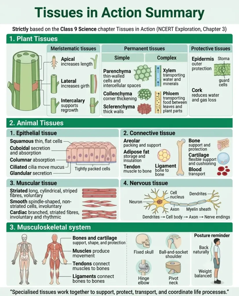
Think about a small seed growing into a tall tree, or a baby growing into a student sitting at a desk. At the start, there is only one cell. That cell divides again and again, and the new cells slowly become skin, muscles, bones, nerves and every other part. How do they know what to become? That question is the heart of this chapter.
In Chapter 2, Cell: The Building Block of Life, you learnt that the cell is the basic unit of life. In a unicellular organism such as Amoeba, one cell does everything: it eats, breathes, moves and reproduces. A large multicellular organism like an elephant or a mango tree cannot work like that. So the work is shared. This sharing of work is called division of labour.
Definition (as in NCERT): A tissue is a group of cells (similar in structure) that work together to perform a specific function.
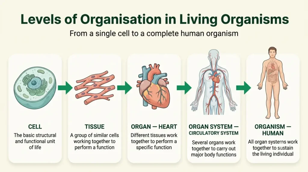
Muscle cells, for example, join together to form muscle tissue, and its job is movement. Different tissues then combine to form an organ. The levels of organisation in a multicellular organism go in this order:
Cell → Tissue → Organ → Organ System → Organism
Let us follow one example from the human body:
- Cell – a single cardiac (heart) muscle cell.
- Tissue – many cardiac muscle cells together form cardiac muscle tissue.
- Organ – the heart. It is not made of muscle alone. It also has connective tissue, epithelial tissue (the lining) and nervous tissue. More than one type of tissue forms an organ.
- Organ system – the heart, blood vessels and blood together form the circulatory system.
- Organism – many organ systems working together form a human being.
Why are tissues important?
- Cells doing the same job work like a team, so the work is done faster and better.
- Division of labour lets the body carry out many complex life processes at the same time.
- Different tissues exist for different jobs. In animals, muscle tissue gives movement and nervous tissue carries messages. In plants, xylem carries water and minerals while phloem carries food.
- The structure of a tissue suits its function. This single idea will help you answer many questions in this chapter. Ask yourself every time: “Why is this tissue built like this?”
Exam point: In multicellular organisms, division of labour is possible because of tissues, and it increases the efficiency of the organism.
Common mistake: Writing that “many similar cells form an organ”. Similar cells form a tissue. An organ needs more than one type of tissue.
Why Are Plant and Animal Tissues Different?
Before we study each tissue, here is a question worth thinking about: why should a plant and an animal have different tissues at all? The answer lies in how they live.
- Most plants are fixed in one place. They need support to stay upright, and plant cells have a rigid cell wall that gives strength.
- Most animals move. Animal cells have no cell wall, so they can change shape easily, and this flexibility helps in locomotion. (A few animals, such as sponges, do not move.)
- Plants make food by photosynthesis, so they have tissues that use sunlight. Animals take in food and need tissues that digest it.
- Both need to carry food and water around the body, but they use different transport tissues: xylem and phloem in plants, and blood in animals.
- The tissues responsible for growth also differ in structure and function. In plants, growth is limited to special growth zones called meristems.
| Feature | Plant tissues | Animal tissues |
|---|---|---|
| Movement | Mostly fixed in one place; need strong support | Most animals move; need flexible cells |
| Cell wall | Present, rigid | Absent |
| Nutrition | Tissues that carry out photosynthesis | Tissues that digest food |
| Transport | Xylem (water, minerals) and phloem (food) | Blood (food, gases, waste) |
| Growth | Confined to meristems (growth zones) | Spread more widely over the body |
| Main groups | Meristematic and permanent | Epithelial, connective, muscular, nervous |
Notice this carefully: The key reason is the lifestyle. Plants stay in one place and need rigid support; animals move and need flexible bodies. When a question asks “why”, always start with this idea.
Plant Tissues
Meristematic Tissues
Look at a growing plant. Its roots go deeper into the soil, its shoot rises upward, and its stem gets thicker year after year. Does growth happen everywhere in the plant? No. It happens only in certain regions where cells can divide. These regions contain meristematic tissue.
Definition: Meristematic tissue is a tissue made of actively dividing cells. It produces new cells continuously and is responsible for the growth of the plant.
Characteristics to learn well:
- Cells are small, closely packed and thin-walled, often roughly equal in all directions (isodiametric).
- They have dense cytoplasm and a large, prominent nucleus.
- They have no vacuole or only very small vacuoles.
- They are living and actively dividing.
- There are no intercellular spaces between them.
Notice this carefully: Every one of these features suits a cell that divides again and again. Dense cytoplasm and a big nucleus support division, and the lack of big vacuoles leaves room for new material.
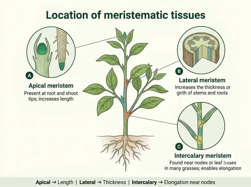
Plants grow in three ways: in length, in girth (thickness) and by regrowth after cutting or grazing. Each kind of growth is done by a different meristem. Based on location, there are three types.
1. Apical meristem – how plants grow in length
Apical meristems are present at the growing tips of roots and shoots. They make the plant longer: taller stems and deeper roots.
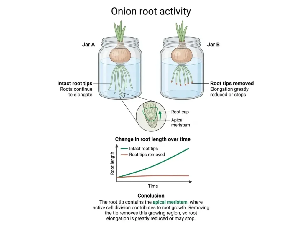
You can prove this yourself with a simple activity from the textbook (Activity 3.1). Keep two onion bulbs, each over a jar of water so that the base touches the water. Measure the length of the roots on the first three days. On day 3, cut about 1 cm from the root tips of the bulb in Jar B and leave Jar A untouched. Keep measuring for four more days and record the lengths in a table.
- Roots in Jar A keep growing in length.
- Roots in Jar B stop growing in length after the tips are cut.
What do you infer? Roots grow in length only from their tips, because the tip contains the dividing cells (the apical meristem). When you remove the tip, you remove the growth zone. You also saw dividing cells in onion root tips when you studied mitosis in the Cell chapter. Shoot tips work in the same way.
2. Lateral meristem – how plants grow in girth
In dicot plants, stems and roots also become thicker with time. This increase in thickness is done by the lateral meristem, which lies along the sides (circumference) of the stem and root, like a ring. The vascular cambium and the cork cambium are lateral meristems.
If you have seen a cut tree trunk in a timber yard, you will have noticed ring-like patterns on the cut surface. These are annual growth rings. Some rings are wide and some are narrow. A wide ring means the tree grew well that year (favourable conditions); a narrow ring means growth was poor. By counting the rings, scientists can estimate the age of the tree and learn about the climate in which it grew.
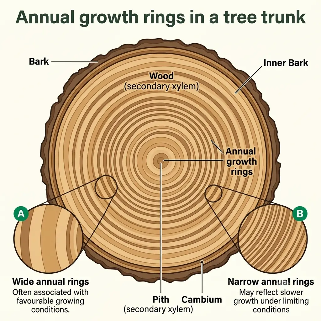
Solved Example – Reading annual rings (illustrative numbers made for practice)
- Given: A cut tree trunk has 22 annual rings and a diameter of 44 cm.
- To find: (i) Approximate age of the tree, (ii) average increase in diameter per year.
- Formula: Age (in years) ≈ number of rings, because one ring forms each year. Average increase = total diameter ÷ number of years.
- Solution: (i) Age ≈ 22 years. (ii) 44 cm ÷ 22 years = 2 cm per year.
- Answer: The tree is about 22 years old, and its diameter increased by about 2 cm per year on average. The real growth was not exactly the same every year, because wide and narrow rings show good and poor years.
3. Intercalary meristem – how plants regrow
Intercalary meristem is present at the nodes, or at the base of internodes (and of leaves in grasses). When grass is mowed or eaten by grazing animals, it grows back, because the intercalary meristem near the base is still there and keeps dividing.
| Type of meristem | Location | Growth it causes | Everyday example |
|---|---|---|---|
| Apical | Tips of roots and shoots | Increase in length | A sapling becoming taller; roots going deeper |
| Lateral | Along the sides of stem and root | Increase in girth | A tree trunk becoming thicker; annual rings |
| Intercalary | Nodes / base of internodes | Regrowth after cutting or grazing | Lawn grass growing back after mowing |
Exam point: Growth in plants takes place only in the meristematic regions. Apical meristem = length, lateral meristem = girth, intercalary meristem = regrowth.
Common mistakes:
- Writing that meristems are found only at the tips. Tips explain length only. Girth and regrowth need lateral and intercalary meristems.
- Writing that intercalary meristem “helps in growth of branches”. It helps in regrowth and growth in length at the nodes/internodes, not in branching.
Permanent Tissues
What happens to the new cells that the meristem makes? They do not keep dividing forever. After some time, they take up a definite shape, size and job. This process is called differentiation. Once differentiated, a cell generally loses the ability to divide and becomes part of a permanent tissue. Some new cells, however, remain meristematic, so the growth zone is never used up.
Definition: Permanent tissues are tissues made of cells that have differentiated from meristematic cells and have a definite shape, structure and function. They usually do not divide.
So permanent tissues come from meristematic tissues. Once permanent, cells can be living or dead, thin-walled or thick-walled. Permanent tissues help in protection, support, conduction (transport) and storage of food, and in photosynthesis in green parts.
Permanent tissues are of two kinds:
- Simple permanent tissue – made of only one type of cell.
- Complex permanent tissue – made of more than one type of cell working together.
(There are also protective tissues, such as the epidermis and cork, which you will meet shortly.)
Exam point (1 mark): The process by which meristematic cells take up a permanent shape and function is called differentiation.
| Feature | Meristematic tissue | Permanent tissue |
|---|---|---|
| Ability to divide | Divides actively | Generally does not divide |
| Cell wall | Thin | Thin or thick |
| Cytoplasm and nucleus | Dense cytoplasm, large nucleus | Cytoplasm less dense; nucleus absent in some dead cells |
| Vacuole | Absent or very small | Often large (e.g., in parenchyma) |
| Intercellular spaces | Absent | Present or absent, depending on the tissue |
| Living or dead | Always living | Living or dead |
| Function | Growth | Protection, support, storage, transport |
Simple and Complex Tissues
A. Simple permanent tissue – made of only one type of cell
There are three types: parenchyma, collenchyma and sclerenchyma.
1. Parenchyma – the basic packing tissue
- Cells are living, thin-walled and loosely arranged, with intercellular spaces and usually a large central vacuole.
- Functions: storage of food and water, and filling the spaces between other tissues. When it contains chlorophyll, it is called chlorenchyma and carries out photosynthesis. In aquatic plants, parenchyma has large air spaces and is called aerenchyma; the trapped air helps the plant float and exchange gases.
- Found in: leaves, stems, roots and fruits.
2. Collenchyma – support with flexibility
- Cells are living and elongated, with walls thickened unevenly, mainly at the corners. The thickening is made of cellulose and pectin.
- Very little or no intercellular space.
- Function: mechanical support with flexibility, so that leaves and stems can bend without breaking.
- Found in: leaf stalks (petioles) and just below the epidermis of stems.
3. Sclerenchyma – hard support
- Cells are long, narrow and dead, with very thick walls hardened by lignin, a chemical that makes the wall stiff. There is no intercellular space.
- Function: gives strength and hardness to the plant.
- Found in: stems, leaf veins, the hard covering of seeds and nuts, the husk of coconut, and fibres such as jute.
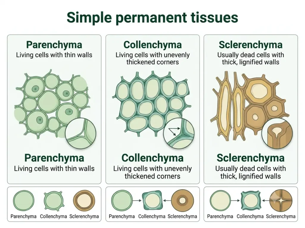
Here is a quick way to remember. Coconut husk fibres are hard because they are sclerenchyma. A coriander leaf stalk bends easily because it has collenchyma. A young sapling bends in strong wind without snapping, and that flexibility also comes from collenchyma.
| Feature | Parenchyma | Collenchyma | Sclerenchyma |
|---|---|---|---|
| Living or dead | Living | Living | Dead |
| Cell wall | Thin | Thickened unevenly at corners (cellulose, pectin) | Very thick, lignified |
| Intercellular spaces | Present | Little or none | None |
| Main function | Storage, photosynthesis (chlorenchyma), floating (aerenchyma) | Flexible support | Hard support, strength |
| Found in | Leaves, stems, roots, fruits | Leaf stalks, below epidermis of stems | Seed coats, coconut husk, fibres |
Common mistake: Saying that all permanent tissues are dead. Parenchyma and collenchyma are living; only some tissues, like sclerenchyma and cork, are dead.
B. Complex permanent tissue – made of more than one type of cell, all working for one function
1. Xylem – conducts water and minerals
- Xylem has four types of elements: tracheids, vessels, xylem parenchyma and xylem fibres.
- Tracheids, vessels and fibres are dead; only xylem parenchyma is living.
- Function: carries water and minerals from the roots to the leaves and other parts, and also gives mechanical support.
- Dead xylem cells form long hollow tubes, which is exactly what is needed for carrying water. In the leaves, water evaporates through the stomata (transpiration). This creates a pulling force, called transpiration pull, that draws water up through the xylem, even against gravity.
2. Phloem – conducts food
- Phloem has four types of elements: sieve tubes, companion cells, phloem parenchyma and phloem fibres.
- Most of these are living; only phloem fibres are dead. Companion cells work alongside the sieve tubes.
- Function: carries food made in the leaves to other parts of the plant, including roots, stems, fruits and growing tips. This movement is called translocation.
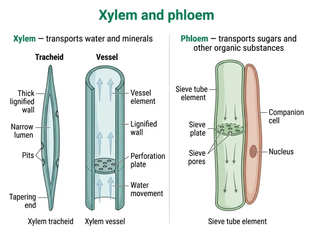
| Xylem | Phloem |
|---|---|
| Conducts water and minerals | Conducts food (made in leaves) |
| Four elements: tracheids, vessels, xylem parenchyma, xylem fibres | Four elements: sieve tubes, companion cells, phloem parenchyma, phloem fibres |
| Mostly dead (only xylem parenchyma is living) | Mostly living (only phloem fibres are dead) |
| Conduction is one-way, upward (roots to leaves) | Conduction is in both directions (up and down) |
| Also gives mechanical support | Does not mainly give support |
Exam point: The definition “a tissue is a group of similar cells” is fully true for simple tissues. In a complex tissue, the cells are of different types, but they still work together for one common function. That is the exact reason why complex tissues are called complex.
Common mistakes:
- Writing that xylem carries food, or that phloem carries water. Check your direction: Xylem = water and minerals (up); Phloem = food (both ways).
- Writing that xylem is made of living cells. Most xylem cells are dead.
Protective Tissues: Epidermis, Stomata and Cork
Every plant needs a “skin” to protect its soft inner parts from injury, germs and loss of water. This is done by the epidermis, and, in old stems, by cork. They are part of the plant’s protective (dermal) tissue.
Epidermis
- It is the outermost layer of cells in the young parts of a plant, such as leaves, flowers, young stems and roots. In most plants, it is usually one cell thick.
- Its cells are flat and tightly packed, with no intercellular spaces.
- It is often covered by a waxy layer called the cuticle, which reduces loss of water. In plants of dry habitats, such as desert plants, the cuticle is thicker. In underwater plants, a thick cuticle would not be useful, because there is no problem of water loss.
- In roots, some epidermal cells have long hair-like extensions that help in absorbing water and minerals.
- The epidermis of leaves has tiny pores called stomata. Each stoma is enclosed by two bean-shaped guard cells, which open and close the pore.
Functions of stomata: (i) exchange of gases (carbon dioxide enters for photosynthesis; oxygen and water vapour go out), and (ii) transpiration, the loss of water vapour, which helps pull water up through the xylem. If a leaf had no stomata, photosynthesis would slow down and transpiration pull would be weak.
Cork
- As a stem gets older, some cells below the epidermis start dividing and form a lateral meristem called cork cambium. It makes new cells on its outer side, and these become cork.
- Cork cells are dead, compactly arranged and have no intercellular spaces. Their walls are heavily thickened with suberin, a waxy chemical that makes them impermeable to water and gases.
- Several layers of cork form the bark of a tree. It protects the inner tissues from injury, germs, water loss and temperature changes, and it replaces the epidermis in old stems.
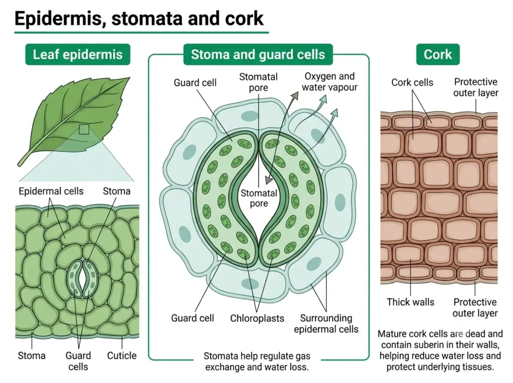
Exam point: Epidermis = protection + reduces water loss (cuticle) + gaseous exchange and transpiration (stomata). Cork = dead, no intercellular spaces, waterproof walls.
The Three Tissue Systems of a Plant
Botanists also group plant tissues by where they lie in the plant body and what they do together.
| Tissue system | Made of | Main work |
|---|---|---|
| Dermal | Epidermis (with cuticle and stomata); cork in older stems | Outer covering; protects inner parts and reduces water loss |
| Ground | Parenchyma, collenchyma, sclerenchyma | Forms much of the plant body between the dermal and vascular tissues; storage, photosynthesis, support |
| Vascular | Xylem and phloem | Transport of water, minerals and food |
Totipotency: When a Permanent Cell Divides Again
We said that permanent cells usually do not divide. But plants hold a surprise. Under the right conditions, even a specialised plant cell can start dividing again and grow into a whole new plant. The ability of a single cell to develop into a complete organism is called totipotency. A fertilised egg (zygote) shows it naturally.
The famous experiment was done by the botanist F. C. Steward and his colleagues in 1958. They took cells from the phloem of a carrot root and grew them in a liquid nutrient medium containing sugars and growth-promoting substances (hormones). The cells lost their specialised features (dedifferentiation), divided and formed a mass of unspecialised cells, and then developed (redifferentiation) into roots, shoots and finally a complete carrot plant.
Why is this useful? The method is the basis of plant tissue culture. It is used for micropropagation (making many identical plants quickly from a tiny piece of a plant) and for producing improved or disease-free plant varieties. Most mature animal cells do not show this kind of totipotency.
Exam point: Totipotency = ability of a cell to grow into a complete organism. Example: a single carrot phloem cell grown in culture (Steward, 1958).
Animal Tissues
Animals move, hunt, think and feel. To do all this, they need tissues that cover, join, contract and carry messages. There are four basic types of animal tissue: epithelial, connective, muscular and nervous.
Epithelial Tissue
Take your skin. It covers your whole body and protects you from dust and germs. The inside of your mouth, food pipe and lungs is also lined by a thin layer. This covering and lining is epithelial tissue.
Definition: Epithelial tissue is the tissue that covers the outer surface of the body and lines the inner surfaces of organs and cavities.
Characteristics:
- Cells are tightly packed with almost no intercellular space and form a continuous sheet.
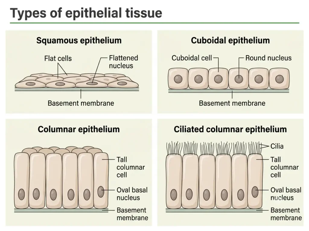
- The sheet rests on a thin non-living layer called the basement membrane.
- It is avascular, which means it has no blood vessels of its own. It gets nutrition by diffusion from the connective tissue below it.
- Its cells are living and divide quickly, so wounds in the skin heal fast.
Types of epithelial tissue (named by shape and arrangement):
- Squamous epithelium – cells are thin, flat and tile-like. A single layer lines the alveoli of the lungs and the blood vessels, where materials must pass quickly. In the skin, the epithelium is stratified (many layers), which protects against wear and tear. Squamous cells also line the mouth and food pipe (oesophagus).
- Cuboidal epithelium – cells are cube-shaped. They help in secretion and absorption. Found in kidney tubules and ducts of salivary glands.
- Columnar epithelium – cells are tall and pillar-like. Found in the inner lining of the small intestine, where it helps in absorption. When the free surface has hair-like cilia, it is called ciliated epithelium. In the respiratory tract, cilia move mucus forward and clear it out.
- Glandular epithelium – some epithelial cells are specialised to secrete substances such as sweat, oil, saliva, mucus and enzymes. They may work singly or form glands (for example, salivary glands and sweat glands).
- Sensory epithelium – specialised receptor cells, often with hair-like projections, that sense the surroundings: smell in the nose, taste in the taste buds, and hearing and balance in the inner ear.
| Function | Structure of epithelium | Where it is found |
|---|---|---|
| Exchange of gases and materials (diffusion) | Single layer of thin, flat cells | Alveoli of lungs, blood vessels |
| Protection | Many layers; outer cells flat and tightly packed | Skin, lining of mouth and oesophagus |
| Secretion | Cuboidal or columnar cells specialised as glands | Salivary glands, sweat glands, stomach lining |
| Absorption | Single layer of tall columnar cells, often with hair-like projections | Lining of small intestine |
| Sensing | Receptor cells, often with cilia | Nostrils, taste buds, inner ear |
Notice this carefully: Epithelium that lines internal organs is usually only one or a few cells thick. A thin sheet allows quick exchange of materials across it. Skin epithelium is thick because its job is protection, not quick exchange. Structure fits function again.
Exam point: Main functions of epithelial tissue: protection, absorption, secretion, exchange of gases and sensing (in some organs, filtration as well).
Common mistake: Writing that blood vessels run inside the epithelium. Epithelium is avascular.
Connective Tissue
If epithelial tissue is the covering, connective tissue is the joining, packing and supporting tissue. It connects organs, supports the body and transports materials.
Definition: Connective tissue is the tissue that connects, supports and binds different tissues and organs of the body.
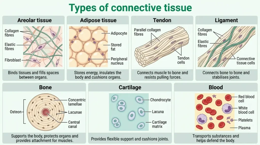
Its cells are loosely spaced and embedded in a non-living matrix. The matrix may be jelly-like, fluid, soft or hard. The type of matrix decides what the tissue can do.
1. Areolar tissue – a loose connective tissue. It fills the spaces inside organs, acts as packing, supports internal organs and helps in the repair of tissues. Found between the skin and muscles, and around blood vessels and organs.
2. Adipose tissue – stores fat. Its cells are filled with fat globules. Found below the skin and around organs such as the kidneys. It acts as an insulator, reduces heat loss and stores energy.
3. Dense regular connective tissue – has fibres and fibre-making cells (fibroblasts) packed in neat rows.
- Tendon – connects muscle to bone. It is very strong, with limited flexibility.
- Ligament – connects bone to bone. It is strong and elastic, holds the joint together, limits movement and helps to prevent dislocation.
4. Skeletal tissue – for support.
- Bone – hard tissue. Its matrix is hard because of calcium and phosphorus salts. It gives shape and a framework to the body and protects organs.
- Cartilage – softer and flexible. Its matrix contains proteins and sugars. Found in the nose, ear and windpipe (trachea), and at the ends of bones in joints. It gives flexibility and absorbs shock.
5. Fluid tissue – for transport.
- Blood – a fluid connective tissue. The matrix is plasma. It contains red blood cells (RBC), white blood cells (WBC) and platelets. RBCs contain haemoglobin, an iron-containing protein that makes blood red and carries oxygen, and they live for about 120 days. WBCs fight germs and gather at the site of infection (this causes swelling, redness and sometimes pus). Platelets help the blood to clot. Blood carries food, gases, hormones and waste.
- Lymph – a colourless fluid made from the liquid part of blood. It carries digested food and waste and helps fight infection. Unlike blood, it has no red blood cells.
You can feel the difference on your own body. Touch your elbow: it is hard and rigid, and that is bone. Press the tip of your ear or nose: it is soft and springs back, and that is cartilage. Wiggle your fingers and feel the forearm: the muscle movement pulls on a tendon. A ligament holds the bones at a joint together and limits the movement.
| Tissue | Matrix | Main function | Where found |
|---|---|---|---|
| Areolar | Soft, jelly-like | Packing, support, repair | Between skin and muscles, around organs |
| Adipose | Soft, with fat-filled cells | Stores fat, insulation | Below skin, around organs |
| Tendon | Dense fibres | Joins muscle to bone | Ends of muscles |
| Ligament | Dense elastic fibres | Joins bone to bone | At joints |
| Bone | Hard (calcium, phosphorus salts) | Support, shape, protection | Skeleton |
| Cartilage | Firm but flexible (proteins, sugars) | Flexibility, cushioning | Nose, ear, windpipe, joints |
| Blood | Fluid (plasma) | Transport | Blood vessels, heart |
Common mistakes:
- Mixing up tendon and ligament. Memory trick: Tendon = To muscle (muscle to bone); Ligament = Links bone to bone.
- Thinking that blood is not a tissue. It is a connective tissue because its cells lie in a fluid matrix (plasma).
Muscular Tissue
Can you move your hand? Does your heart beat? All this happens because of muscular tissue.
Definition: Muscular tissue is made of long cells called muscle fibres. They contain special contractile proteins, which contract and relax and cause movement.
There are three types.
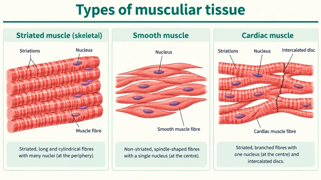
1. Striated (skeletal or voluntary) muscle
- Cells are long, cylindrical, unbranched and multinucleate (many nuclei). Under the microscope, they show alternate light and dark bands called striations.
- They are voluntary: you can move them by your wish. Example: muscles of the arms and legs.
- Usually attached to bones by tendons. They get tired with prolonged work.
2. Unstriated (smooth or involuntary) muscle
- Cells are long, spindle-shaped, with pointed ends and a single nucleus. There are no striations.
- They are involuntary: you cannot control them by will. Found in the walls of the stomach, intestine and blood vessels.
- They work slowly and do not get tired easily.
3. Cardiac muscle – found only in the heart
- Cells are cylindrical and branched, usually with a single nucleus (sometimes two). They are also striated, but the striations are less regular-looking than in skeletal muscle.
- They are involuntary and contract rhythmically throughout life. Under normal conditions, they do not show fatigue the way skeletal muscle does.
- The ends of neighbouring cells are joined by intercalated discs, which allow the heart cells to contract together in step.
| Feature | Striated (skeletal) | Unstriated (smooth) | Cardiac |
|---|---|---|---|
| Control | Voluntary | Involuntary | Involuntary |
| Cell shape | Long, cylindrical, unbranched | Spindle-shaped, pointed ends | Cylindrical, branched |
| Nuclei | Many (multinucleate) | One | Usually one |
| Striations | Present, clear | Absent | Present, less regular |
| Special feature | Attached to bones | Slow, sustained contraction | Intercalated discs |
| Found in | Limbs, attached to bones | Stomach, intestine, blood vessels | Heart only |
Exam point: The heart is made of cardiac muscle, which is involuntary and works without rest throughout life.
Common mistake: Writing that all involuntary muscles are smooth. Cardiac muscle is also involuntary, but it is striated and branched.
Nervous Tissue
How do you feel hot and cold? How does your brain tell your hand to write? This very fast communication is done by nervous tissue.
Definition: Nervous tissue is the tissue that controls and coordinates the activities of the body by carrying messages in the form of electrical signals called nerve impulses.
It contains two kinds of cells: neurons and supporting cells called neuroglia.
Neuron is the structural and functional unit of nervous tissue. It is one of the longest cells in the body; some neurons that run from the spinal cord to the foot are about a metre long. Not every neuron is that long, though.
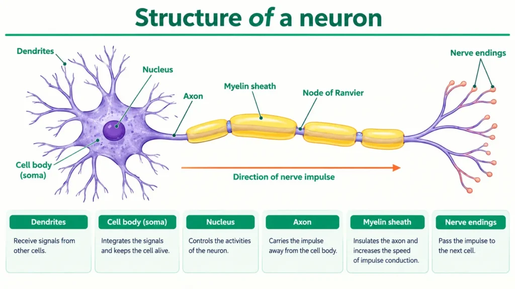
Structure of a neuron (you may be asked to draw it):
- Cell body (cyton) – contains the nucleus and cytoplasm.
- Dendrites – many short, branched fibres from the cell body. They receive the impulse.
- Axon – one long fibre that carries the impulse away from the cell body to the next cell. In many neurons, it is covered by a fatty myelin sheath, which helps fast conduction. The axon ends in branched nerve endings.
Functions:
- It receives stimuli from sense organs such as the eyes and ears.
- It carries messages as nerve impulses to the brain, spinal cord and muscles.
- It helps the body respond quickly to changes in the surroundings.
Exam point: Nervous tissue can be excited and can conduct impulses. That is why it controls and coordinates the body.
Animal Tissues at a Glance
| Tissue | Cells and matrix | Main function | Examples |
|---|---|---|---|
| Epithelial | Tightly packed cells, almost no matrix; basement membrane | Covering, lining, protection, absorption, secretion, exchange, sensing | Skin, lining of mouth, lungs, intestine |
| Connective | Loosely spaced cells in a matrix | Connect, support, bind, transport, store fat | Areolar, adipose, tendon, ligament, bone, cartilage, blood |
| Muscular | Long contractile fibres | Movement | Skeletal, smooth, cardiac muscle |
| Nervous | Neurons and neuroglia | Control and coordination | Brain, spinal cord, nerves |
Musculoskeletal System
Elementary Idea and Joints
You can stand straight, walk, run and hold a pen. This is possible because of a framework of bones and muscles working together. This is called the musculoskeletal system.
Definition: The musculoskeletal system is made of the skeleton (bones and cartilage), muscles and joints. It gives shape, supports the body, protects organs and helps in movement.
The skeleton and muscles – an elementary idea
- The adult human skeleton typically has 206 bones, along with cartilage. (A baby is born with more than this; many bones fuse as the child grows.)
- The skeleton gives the body shape and a framework. The skull protects the brain, the rib cage protects the heart and lungs, and the vertebral column (backbone) supports the body and protects the spinal cord.
- Muscles are attached to bones by tendons, and bones are joined to bones by ligaments. When a muscle contracts, it pulls the bone, and the bone moves at a joint.
- Movement is controlled by the nervous system, which sends signals to the muscles.
Joints
Definition: A joint is a place where two or more bones meet.
Joints are of different kinds, depending on how much movement they allow.
1. Fixed (immovable) joint – no movement. Example: the joints between most bones of the skull (the lower jaw is the exception, because it moves). This keeps the brain safe.
2. Freely movable (synovial) joints – allow a lot of movement. They have a space filled with synovial fluid, which acts as a lubricant and reduces friction, and cartilage at the bone ends that works as a cushion. The main kinds are:
- Ball and socket joint – allows movement in all directions. Example: shoulder and hip.
- Hinge joint – allows bending and straightening in one plane, like a door hinge. Example: elbow and knee (the ankle also moves in a hinge-like way).
- Pivot joint – allows rotation. Example: neck, where the first vertebra turns on the second (this lets you shake your head to say “no”).
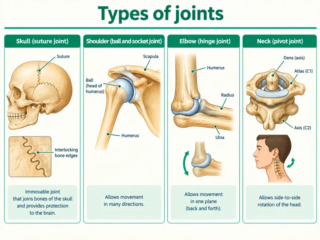
Extra detail (only if your teacher covers it): Some books also describe a gliding joint (bones slide over each other, as in the wrist and ankle) and a slightly movable (cartilaginous) joint (for example, between the vertebrae of the backbone, where cartilage lies between the bones). The textbook focuses on the four types above, so write these two only if asked.
| Joint | Movement | Example |
|---|---|---|
| Fixed | None | Skull bones |
| Ball and socket | In all directions | Shoulder, hip |
| Hinge | Bend and straighten (one plane) | Elbow, knee |
| Pivot | Rotation | Neck (atlas on axis) |
Try this at home: Jump normally and notice how your ankles, knees and hips bend before the push and straighten afterwards. Now try a jump with stiff legs: the jump is much weaker. Bending at the joints lets the muscles store and release force.
Notice this carefully: Synovial joints allow a wide range of movement because of the way their bone ends fit together and their joint cavity. The synovial fluid reduces friction, so the movement is smooth. It does not create the range of movement; it makes movement smooth.
Exam point: Ball and socket joints give the maximum range of movement. Hinge joints work in one plane. Pivot joints allow rotation.
How Tissues Work Together in Movement
Think of bending your arm to lift a bag. Many tissues team up:
- Nervous tissue sends the signal (“lift!”) from the brain to the muscle.
- Muscular tissue contracts and pulls.
- Tendon (connective tissue) transmits the pull from the muscle to the bone.
- Bone acts as a rigid lever that moves around the joint.
- At the joint, cartilage cushions the bone ends, ligaments hold the bones together and synovial fluid reduces friction.
Muscles can only pull, not push. So they often work in pairs: when one contracts, the other relaxes, and this lets the joint bend one way and straighten the other way. The same teamwork happens in dancing, yoga and sports. A dancer’s smooth poses, a cricketer’s throw and a footballer’s kick all depend on well-coordinated muscles, bones, joints and nerves.
Care: Injuries, Postural Care, Nutrition and Exercise
Bones and muscles stay healthy only if we look after them. Otherwise, injuries and pain follow.
A. Common injuries
| Injury | What is hurt | Usual cause | What to do |
|---|---|---|---|
| Sprain | Ligament (stretched or torn) | Twisting or overstretching a joint; common in the ankle | Protect and support the joint; see a doctor if pain or swelling is severe |
| Strain | Muscle or tendon | Overstretching or overuse | Rest the muscle, return to activity gradually |
| Fracture | Bone (cracked or broken) | Fall, accident or strong blow | Keep the part still and supported and get medical help; the doctor may use a splint or plaster cast |
| Dislocation | Bone slips out of its joint (e.g., shoulder) | Strong pull or fall | Keep the joint still and get medical help; do not try to push the bone back yourself |
RICE: Many textbooks teach Rest, Ice, Compression and Elevation as the first-aid steps for minor sprains and strains, so you should know it for your exam. Doctors, however, now debate it. Newer advice (called “PEACE & LOVE”, proposed by physiotherapists in 2019–2020) says ice only gives short-term pain relief, and that too much rest can delay healing, so gentle movement within pain limits is encouraged. In real life, follow your doctor’s advice, and see a doctor at once if you cannot put weight on the limb or the shape looks wrong.
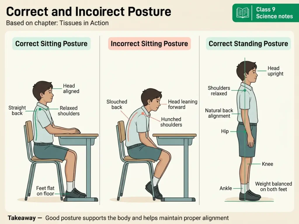
B. Postural care – correct posture
Definition: Posture is the position of the body while sitting, standing and walking.
- Keep your back straight while sitting. Do not slouch.
- While standing, put your weight equally on both legs.
- Use a chair with back support and a table of the right height while studying.
- Do not carry a heavy school bag on one shoulder. Use both straps, so that the load is shared; a bad habit can strain the back and spine over time.
C. Nutrition for bones and muscles
- Calcium – for strong bones and teeth. Sources: milk, curd, cheese, green leafy vegetables.
- Vitamin D – helps the body absorb calcium. Sources: sunlight, eggs, fish.
- Proteins – for building and repairing muscles. Sources: pulses, eggs, chicken.
- Phosphorus – also needed for hard bones.
D. Exercise
- Regular exercise strengthens bones and muscles and keeps joints flexible. Walking, running, yoga and stretching are good examples.
- It also improves blood circulation and helps to prevent obesity.
- Never begin very heavy exercise suddenly. Warm up first.
- If you get a muscle injury, doctors usually advise a short period of protection, then slow and gradual return to activity. Follow the advice of a doctor or physiotherapist for recovery.
Exam point: A balanced diet rich in calcium and vitamin D, correct posture and regular exercise are needed to keep the musculoskeletal system healthy.
Common mistake: Using “sprain” and “strain” as if they mean the same thing. A sprain hurts a ligament; a strain hurts a muscle or tendon.
Related References
Yoga and Physical Agility
You have studied bones, muscles and joints. But how can we keep them flexible and agile? One answer comes from India: yoga. The CBSE syllabus asks you to explain how yoga helps physical agility and correct posture.
Definition: Yoga is an ancient Indian practice that combines body postures (asanas), breathing control (pranayama) and meditation to keep body and mind healthy.
The word yoga comes from the Sanskrit root yuj, which means “to join” or “to unite”, for example, joining body and mind. Physical agility means the ability to move the body quickly and easily with flexibility and balance.
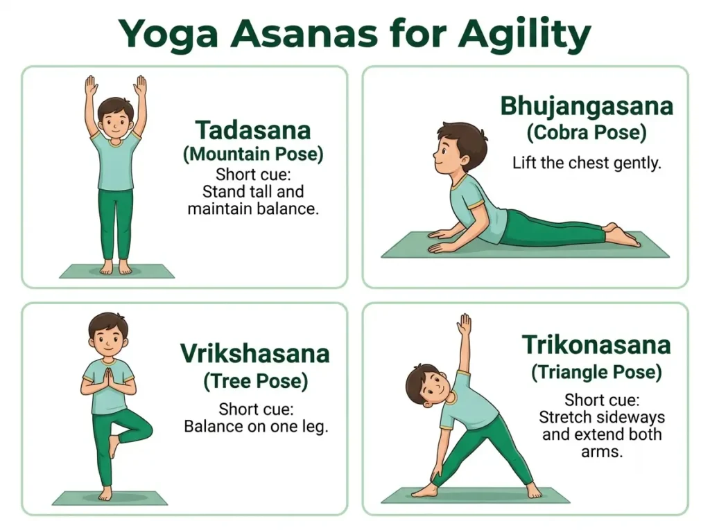
Some asanas that help the musculoskeletal system:
- Tadasana (Mountain Pose) – standing straight with arms raised. It improves posture and balance and stretches the spine.
- Bhujangasana (Cobra Pose) – lying on the stomach and lifting the chest. It makes the backbone flexible and strengthens the back muscles.
- Vrikshasana (Tree Pose) – standing on one leg. It improves balance and concentration and strengthens the legs.
- Trikonasana (Triangle Pose) – stretching the legs and the sides of the body. It improves the flexibility of joints and muscles.
Benefits of regular yoga (write these in your answer):
- It increases the flexibility and strength of muscles and joints.
- It improves posture and can reduce strain on the back.
- It supports good blood circulation and fitness.
- It reduces stress and improves concentration, which helps in studies too.
- It is low-cost and can be practised at home.
Safety note: Yoga is generally safe when learnt properly, but forcing a posture can cause injury. Learn from a trained teacher, warm up first, never push into pain, and ask a doctor first if you have an injury or a health problem.
Exam point: Regular yoga practice keeps the musculoskeletal system flexible and agile and helps maintain the correct posture.
Indian Contributions
Your syllabus asks you to appreciate India’s contributions to science, with examples. For this chapter, it names two scientists of plant cell and tissue culture. Ancient Indian texts on the body and on yoga also give useful background.
1. Prof. Sipra Guha Mukherjee and Prof. S. C. Maheshwari – plant tissue culture
In 1964, Sipra Guha and S. C. Maheshwari reported that when anthers (the pollen-bearing parts of a flower) of the thorn-apple plant Datura were grown on a nutrient medium in the laboratory, the pollen grains developed into embryos and then into plants with only one set of chromosomes (haploid plants). This technique, called anther culture, became widely used in plant breeding. It shows the idea you met in totipotency: plant cells, grown in the right conditions, can form whole plants.
2. Patanjali and the Yoga Sutras
Patanjali is credited with compiling the Yoga Sutras, a collection of short sayings (sutras) on yoga. He organised older ideas rather than inventing yoga from nothing. The text describes the eight limbs of yoga (Ashtanga yoga): yama, niyama, asana, pranayama, pratyahara, dharana, dhyana and samadhi. Scholars do not agree on the date of the text; estimates range from about the 2nd century BCE to the 4th–5th century CE, so “about 2000 years ago” is only a rough statement.
3. Sushruta and the Sushruta Samhita
The Sushruta Samhita is an ancient Indian text on medicine and surgery. Sushruta is often called the “Father of Surgery”. The text supports learning anatomy by dissection, and it describes more than 300 bones along with joints, ligaments and muscles. It also discusses fractures and dislocations and their treatment. A date of about 600 BCE is often quoted, but it is an estimate; scholars place the work anywhere from the first millennium BCE to the early centuries CE, and the surviving text was revised over time.
4. Charaka Samhita and Ayurveda
The Charaka Samhita is another major ancient text of Ayurvedic medicine, and it includes a section on the anatomy of the body. In Ayurveda, bone is asthi, a joint is sandhi and muscle (flesh) is mamsa.
| Name | Known for | What to remember |
|---|---|---|
| Guha Mukherjee and Maheshwari | Anther culture in Datura (1964) | Plant tissue culture research in India |
| Patanjali | Yoga Sutras; eight limbs of yoga | Date uncertain; he compiled older traditions |
| Sushruta | Sushruta Samhita; surgery and anatomy | Described bones, joints, muscles; date uncertain |
| Charaka | Charaka Samhita; medicine | Major text of Ayurveda |
Common mistake: Writing exact dates for Patanjali or Sushruta as if they were certain. Use words like “traditionally” or “about”, and do not give a date unless your teacher does.
Exam point: India’s contributions you can quote: yoga for agility and posture, the Sushruta Samhita for early knowledge of bones, joints and surgery, and the work on plant tissue culture by Sipra Guha Mukherjee and S. C. Maheshwari.
Practical: Identifying Tissues from Prepared Slides (Experiment 5)
The CBSE list of practicals includes this experiment for Chapter 3. Practical work carries 5 marks (Subject Enrichment) in the 20-mark internal assessment, shared across all your practicals.
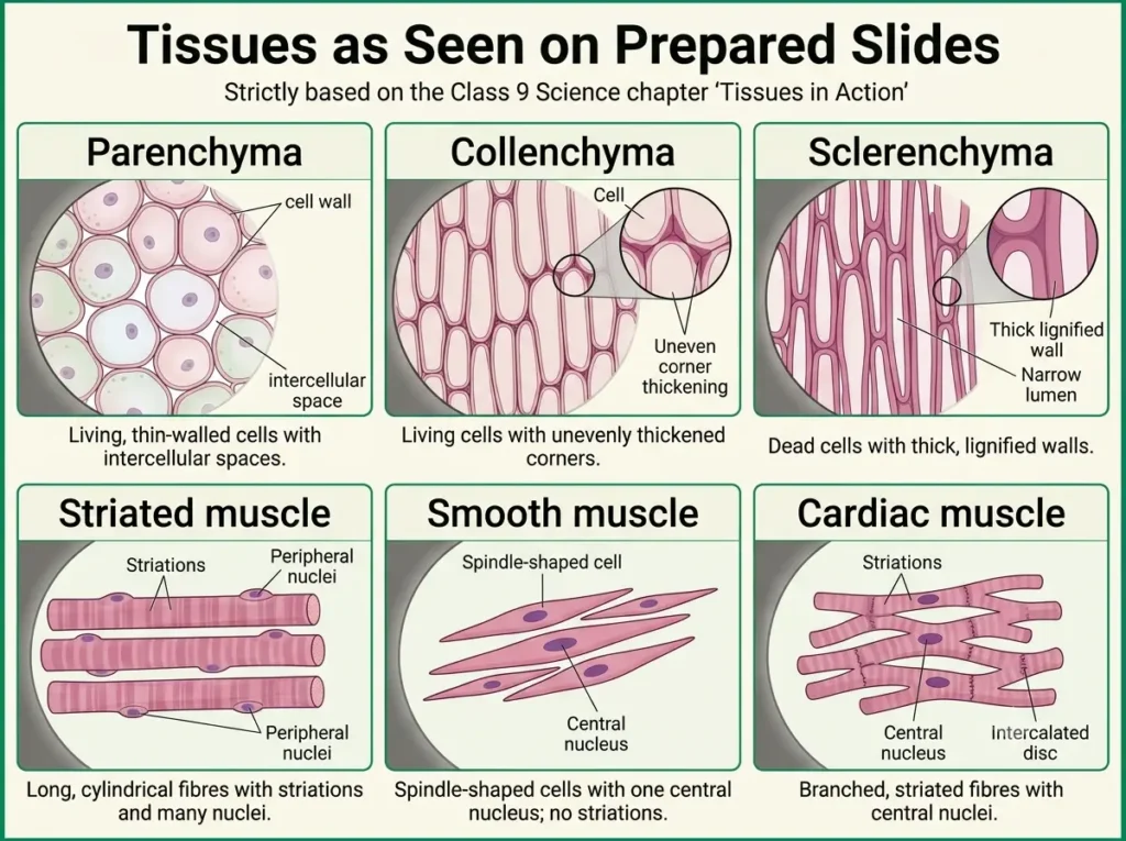
Aim: To study and identify tissues in plants (parenchyma, collenchyma and sclerenchyma) and in animals (striped, smooth and cardiac muscle fibres) from prepared slides, and to draw their labelled diagrams.
Materials required: Compound microscope, prepared permanent slides of parenchyma, collenchyma, sclerenchyma, striated muscle, smooth muscle and cardiac muscle, practical notebook, pencil.
Procedure:
- Place a prepared slide on the stage of the microscope and fix it with the clips.
- First observe under the low-power objective (usually 10X).
- Use the coarse adjustment to find the object, and then the fine adjustment to make it sharp.
- Switch to the high-power objective (usually 40X; some school microscopes have 45X) only after the object is clear under low power.
- Draw what you see and label the important parts. Repeat for every slide.
Observation – how to identify each tissue:
| Slide | What you will see | Identification | Function |
|---|---|---|---|
| Parenchyma | Thin-walled, living, roundish or oval cells with intercellular spaces and a large vacuole | Simple permanent tissue | Storage, filling |
| Collenchyma | Living, elongated cells with thickening at the corners; very little intercellular space | Simple permanent tissue | Flexible support |
| Sclerenchyma | Dead cells with very thick lignified walls, narrow cavity, no intercellular space | Simple permanent tissue | Hard support, strength |
| Striated (skeletal) muscle | Long cylindrical fibres, unbranched, light and dark bands, many nuclei | Voluntary muscle | Movement of limbs |
| Smooth muscle | Spindle-shaped cells with pointed ends, one central nucleus, no striations | Involuntary muscle | Movement in internal organs |
| Cardiac muscle | Branched fibres, striations, usually one nucleus, intercalated discs | Involuntary muscle | Heartbeat |
Extra: Some schools also show a prepared slide of a neuron (nerve cell), with its cell body, many short dendrites and one long axon. A neuron slide is not part of the CBSE list for this experiment, so draw it only if your teacher asks.
Precautions:
- Handle the microscope and slides carefully.
- Do not touch the cover slip of a prepared slide.
- Always start from low power and then go to high power.
- Draw neat, clear and labelled diagrams.
Viva voce – quick answers:
- Which of the three plant tissues is dead? Sclerenchyma.
- Why is sclerenchyma strong? Its walls are very thick and hardened with lignin.
- Which muscle has intercalated discs? Cardiac muscle.
- Which muscle is voluntary and multinucleate? Striated (skeletal) muscle.
Related activity (apical meristem): The syllabus also lists an experiment to understand plant growth due to the apical meristem. The onion-root activity described under Meristematic Tissues is the one to do.
Conclusion
Tissues are where the idea of division of labour becomes real. A plant grows because its meristems keep dividing, and it stays strong, protected and supplied because its permanent tissues each do one part of the job: protection from the epidermis and cork, storage and photosynthesis in parenchyma, flexible support in collenchyma, hard strength in sclerenchyma, and transport through xylem and phloem. In animals, epithelial tissue covers and lines, connective tissue joins and supports, muscular tissue moves, and nervous tissue controls. Bones, muscles, tendons, ligaments and joints work as one musculoskeletal system, kept healthy by good food, correct posture, exercise and, as our tradition teaches, yoga. As you revise, keep asking the same question for every tissue: how does its structure suit its function?
Quick Summary
- Tissue and organisation: A tissue is a group of similar cells doing a specific job. Cell → tissue → organ → organ system → organism. Division of labour makes multicellular organisms efficient.
- Plant tissues: Meristematic tissues (apical = length, lateral = girth, intercalary = regrowth) divide actively. Permanent tissues are simple (parenchyma, collenchyma, sclerenchyma) or complex (xylem carries water and minerals up; phloem carries food in both directions). The epidermis and cork protect the plant.
- Animal tissues: Epithelial (covers and lines, avascular), connective (matrix; blood, bone, cartilage, tendon, ligament), muscular (striated, smooth, cardiac) and nervous (neurons).
- Movement and care: Muscles pull bones through tendons; ligaments join bone to bone; joints are fixed, ball and socket, hinge or pivot. Calcium, vitamin D, proteins, posture, exercise and yoga keep the system healthy.
Previous Year and Practice Questions
Class 9 Science Ch 3 – Tissues Previous Year Questions
Frequently asked questions from CBSE / NCERT Exemplar papers on Tissues. Tap a question to view a quick-answer guide.
1 The husk of coconut is made up of which tissue? +
2 Which chemical makes cork cells impervious to water and gases? +
3 Name the tissue responsible for flexibility in plants. Where is it located? +
4 What is the basic principle for categorising plant tissues as meristematic and permanent? +
5 A permanent slide shows thin-walled isodiametric cells with large vacuole. Identify the tissue. [CBSE 2011] +
6 Differentiate between meristematic and permanent tissues. +
7 Why are xylem and phloem called complex tissues? How are they different? +
8 Why do plants have more dead tissues compared to animals? +
9 Tissue A and Tissue B constitute Tissue C. A carries water while B carries food. Identify A, B, C. +
10 The root tips of a plant were cut and the plant was replanted. What will happen and why? +
11 Explain location and function of three types of meristematic tissue. +
12 Water hyacinth floats on water surface. Explain. +
13 What are functions of stomata? +
14 Differentiate between parenchyma and sclerenchyma. +
15 List characteristics of cork. How are they formed? Mention their role. +
16 Why are plants and animals made of different types of tissues? +
17 Differentiate between tendon and ligament. +
18 What are components of blood? Why is blood considered connective tissue? +
19 Give three features of cardiac muscles. +
20 Identify the tissues: (a) inner lining of mouth (b) connects muscle to bone (c) transports food in plants (d) stores fat +
Frequently Asked Questions
Is “Tissues in Action” the same as the old Chapter 6, “Tissues”?
It covers the same core idea, but it is a new chapter in the new Exploration book. It is now Chapter 3, and it adds activities and topics such as totipotency and the musculoskeletal system. Treat older notes as a rough guide only, and follow the NCERT book.
If xylem cells are dead, how do they carry water upward?
Dead cells form long, hollow tubes, and a tube needs no living contents to carry water. The pull comes from the leaves, where water evaporates through the stomata (transpiration pull), and the living cells of the leaf keep this process going.
Why do roots stop growing in length when the tip is cut?
Cell division for length happens in the apical meristem at the tip. Cutting the tip removes it. This is the idea behind the onion-root activity.
How can I remember tendon and ligament?
Tendon = To muscle (muscle to bone). Ligament = Links bone to bone. A sprain hurts a ligament; a strain hurts a muscle or tendon.
Why does the heart not get tired like other muscles?
Under normal conditions, cardiac muscle does not fatigue the way skeletal muscle does. It is rich in mitochondria and has a good blood supply, and its cells are joined by intercalated discs so that they contract together in a steady rhythm.
Why is blood called a tissue?
Because it fits the definition of a connective tissue: its cells (RBC, WBC, platelets) lie in a non-living matrix, the fluid plasma.
Which joint allows the maximum movement?
The ball and socket joint (shoulder and hip). The shape of its bone ends allows movement in all directions, and synovial fluid keeps the motion smooth.
Should I always use ice and complete rest after a sprain?
RICE is the first-aid method taught in most schools, so write it in exams. Newer medical advice questions long periods of rest and routine icing, and encourages gentle movement as pain allows. For a real injury, ask a doctor.
Continue Reading
Continue Reading – Class 9 Science (NCERT Exploration)
- Cell: The Building Block of Life – Class 9 Science Notes (Chapter 2) – revise the cell, organelles, osmosis and cell division before this chapter.
External References
Continue Reading — Class 9 Science: Tissues in Action
Subscribe to keep reading
You've used your free notes for now. Get Delhi Pathshala Premium for unlimited access.
Subscribe — ₹29/30 days