
Contents
- What You Will Learn
- Key Terms at a Glance
- Cell: Introduction
- Discovery of Cell
- Cell as Structural and Functional Unit of Life
- Seeing Cells: Microscope, Magnification and Size
- Types of Cells
- Plant and Animal Cells
- Prokaryotic and Eukaryotic Cells
- Cell Organelles
- Nucleus
- Mitochondria
- Chloroplast
- Endoplasmic Reticulum
- Vacuoles
- Plasma Membrane
- Cell Wall
- Ribosomes
- Golgi Apparatus
- Lysosomes
- Cytoplasm and Other Parts
- Permeability of Cell Membranes
- Diffusion and Osmosis
- Cellular Division and Cancer
- Mitosis and Meiosis
- Cancer
- Recent Advancement in Cell Biology
- Case Study and Indian Contribution
- Leigh Syndrome and Mitochondrial Dysfunction
- Contribution of Prof. Arun Kumar Sharma
- Can We Make an Artificial Cell?
- Practical: Preparing Stained Temporary Mounts
- Preparing Stained Temporary Mounts of Onion Peel and Human Cheek Cells
- Conclusion
- Quick Summary
- NCERT-Style Questions with Answer Points
- Frequently Asked Questions
- External References
Quick answer: A cell is the basic structural and functional unit of all living organisms. Every living thing, from a bacterium to a banyan tree, is made of one or more cells, and every life process takes place inside them. These notes cover Chapter 2 of the new NCERT Class 9 Science textbook, Exploration, for the CBSE session 2026-27: the discovery of the cell, plant and animal cells, prokaryotic and eukaryotic cells, all major organelles, osmosis, cell division, cancer, the Leigh syndrome case study, an Indian scientist, and the onion-peel and cheek-cell practical.
Note for students: The 2026-27 book is new. Its Chapter 2 is Cell: The Building Block of Life and it replaces the old Chapter 5, The Fundamental Unit of Life. If your school still uses older notes, the facts are similar, but the chapter number, activities and exercise questions are different. Always follow your own teacher’s instructions and the NCERT textbook.
What You Will Learn
| In this chapter you will learn… | Where to find it |
|---|---|
| Who discovered the cell and how the cell theory developed | Discovery of Cell |
| Why the cell is called the structural and functional unit of life | Cell as Structural and Functional Unit of Life |
| How to estimate the size of a cell and calculate magnification | Seeing Cells: Microscope, Magnification and Size |
| Plant cell vs animal cell; prokaryotic cell vs eukaryotic cell | Types of Cells |
| Structure and function of every key organelle | Cell Organelles |
| Diffusion, osmosis, isotonic, hypotonic and hypertonic solutions | Permeability of Cell Membranes |
| Mitosis, meiosis, the cell cycle and cancer | Cellular Division and Cancer |
| Leigh syndrome, Prof. Arun Kumar Sharma and the artificial-cell question | Recent Advancement in Cell Biology |
| Steps of the onion-peel and cheek-cell practical | Practical |
| Practice questions, case-based questions and FAQs | Practice Questions |
Key Terms at a Glance
| Term | Meaning in one line |
|---|---|
| Cell | The basic structural and functional unit of a living organism |
| Cell theory | All organisms are made of cells; the cell is the basic unit of life; all cells arise from pre-existing cells |
| Plasma (cell) membrane | Thin, flexible, selectively permeable boundary of every cell |
| Cell wall | Rigid, permeable outer layer in plant, fungal and bacterial cells |
| Cytoplasm | Semi-fluid, jelly-like substance inside the cell where organelles lie |
| Nucleus | Organelle that holds the DNA and controls the cell’s activities |
| Nucleoid | Region of a prokaryotic cell where its circular DNA lies without a membrane around it |
| Chromatin / Chromosome | Thread-like DNA–protein material in a non-dividing cell / rod-shaped form seen before division |
| Gene | A functional segment of DNA |
| Organelle | A part inside the cell that does a specific job |
| Mitochondria | Organelles that release energy from food and store it as ATP |
| Chloroplast | Green plastid containing chlorophyll where photosynthesis takes place |
| ER, Golgi, lysosome | Transport and manufacturing network, packaging unit, and clean-up unit of the cell |
| Vacuole | Fluid-filled sac that stores materials and gives support |
| Diffusion | Net movement of particles from higher to lower concentration |
| Osmosis | Movement of water across a selectively permeable membrane |
| Mitosis / Meiosis | Division giving two identical cells / division giving four cells with half the chromosomes |
Cell: Introduction
Discovery of Cell
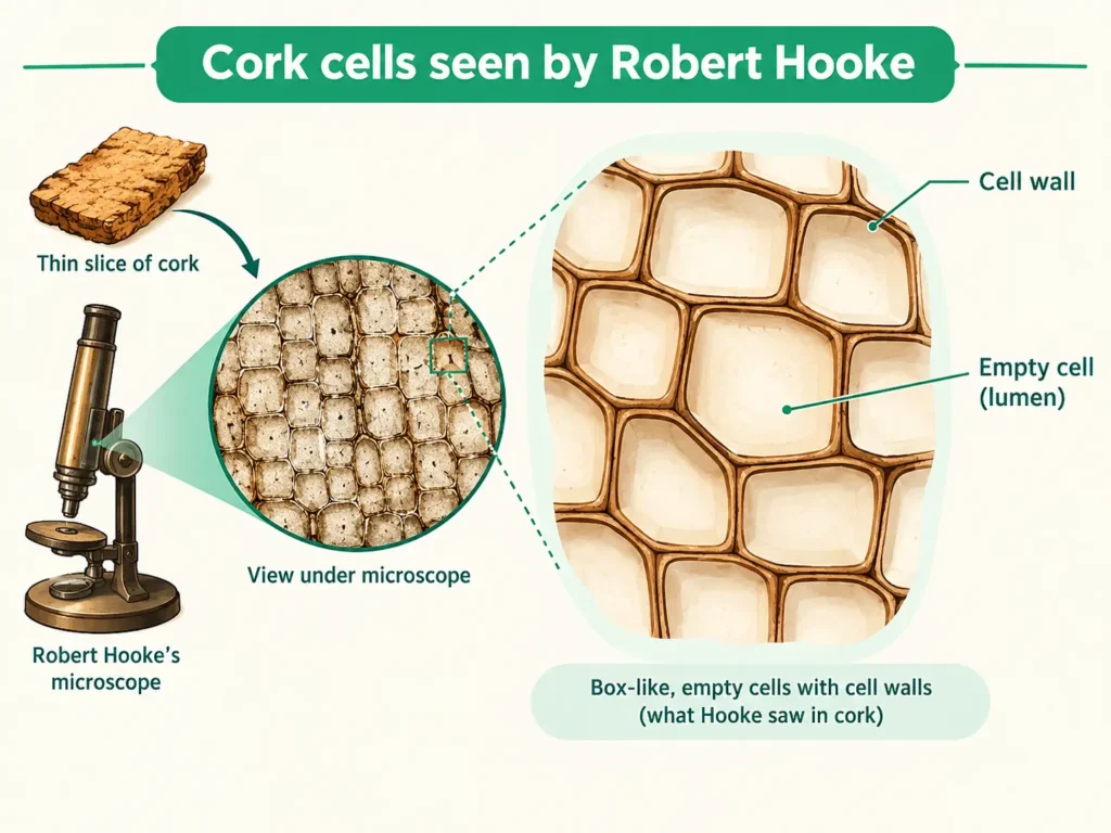
The story of the cell began when people learnt to make better microscopes. In 1665, Robert Hooke looked at a thin slice of cork (a part of tree bark) under a microscope he had designed himself. He saw many small, box-like compartments that reminded him of tiny rooms. He called them cellulae, a Latin word meaning small rooms, and that is where our word “cell” comes from.
Notice this carefully: what Hooke saw were not living cells. Cork is dead tissue, so he was really looking at empty cell walls.
Many scientists then added to the picture. The dates below are the ones usually given in Class 9 books; historians sometimes quote slightly different years, so use the years your textbook gives.
| Year | Scientist | Contribution |
|---|---|---|
| 1665 | Robert Hooke | Observed cork under a self-designed microscope and named the “cells” |
| 1674 | Anton van Leeuwenhoek | Used improved lenses to see living cells such as bacteria and protozoa in pond water; often called the father of microbiology |
| 1831 | Robert Brown | Described the nucleus in the cell (his findings were published in 1833) |
| 1838 | M. J. Schleiden | A botanist; concluded that all plants are made of cells |
| 1839 | T. Schwann | A zoologist; found the same for animals; together with Schleiden he is credited with the cell theory |
| 1839 | J. E. Purkinje | Credited with the term protoplasm for the living substance of the cell |
| 1855 | Rudolf Virchow | Added that all new cells arise from pre-existing cells |
Exam point: The cell theory has three ideas: (i) all living organisms are made of one or more cells; (ii) the cell is the basic structural and functional unit of life; (iii) all cells arise from pre-existing cells. Examiners often ask this point, and the third idea (Virchow’s) is the one students forget.
Common mistake: Writing that Hooke discovered living cells, or that Brown discovered the cell. Hooke discovered cells (dead cork cells); Brown discovered the nucleus.
Cell as Structural and Functional Unit of Life
Think of a building. It is made of thousands of bricks, and each brick is a small unit. In the same way, every living organism is built from cells, so the cell is called the structural unit of life.
Definition: The cell is the structural and functional unit of life. Keep this line ready for one-mark questions.
As a structural unit
- Some organisms, like Amoeba and bacteria, are made of one cell only. They are unicellular.
- Others, like humans, a mango tree or a dog, are made of many cells. They are multicellular. Your body contains trillions of cells.
- Cells differ in shape and size according to their work. A nerve cell is long and thin, a red blood cell is a small disc, and a plant root hair cell has a long extension that helps it absorb water.
- In multicellular organisms, a group of similar cells forms a tissue, tissues form an organ, and organs working together form an organ system. You will study tissues in the next chapter, Tissues in Action.
As a functional unit
All the work of life is done inside cells. When you eat, breathe or grow, it is your cells that do the small jobs that add up to these big functions.
- Respiration, digestion, making new proteins and removing waste all happen inside the cell.
- In a unicellular organism, a single cell does every job.
- In a multicellular organism, different cells do different jobs, but together they keep the organism alive.
So a cell is not just a building block. It is also a working factory.
Seeing Cells: Microscope, Magnification and Size
Why this section matters: The 2026-27 textbook begins the chapter by asking how we can study something so small. These ideas are used in the practical and in numerical questions. If your teacher has not asked you to prepare this part, treat it as supporting reading.
The smallest distance between two points that the human eye can still see as two separate points is its limit of resolution, about 0.1 mm. Most cells are smaller than this, so we need a microscope.
A microscope improves three things:
- Magnification – how many times larger the image looks than the object. The symbol X means “times”, so 400X means 400 times larger.
- Resolution – how clearly two close points can be seen as separate.
- Contrast – the difference in brightness between different parts of the object (staining improves contrast).
School light microscopes use lenses such as 10X (eyepiece) and 40X (objective). Electron microscopes use a beam of electrons instead of light and can show details on the scale of nanometres (1 nm is one-billionth of a metre).
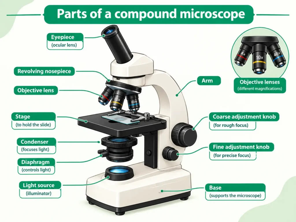
Formulas and units
- Estimated size of a cell = (diameter of the visible field in µm) ÷ (number of cells across the diameter)
- Total magnification = eyepiece magnification × objective magnification
- 1 mm = 1000 µm (micrometres)
Solved Example 1 – Estimating cell size
- Given: Diameter of the visible field = 2 mm; number of cells along the diameter = 25
- To find: Estimated size of one cell
- Formula: Cell size = field diameter (µm) ÷ number of cells
- Solution: Convert first: 2 mm × 1000 = 2000 µm. Then 2000 µm ÷ 25 = 80 µm
- Answer: One cell is about 80 µm long.
Solved Example 2 – Total magnification and apparent size
- Given: Eyepiece = 10X, objective = 40X; real length of a cell = 50 µm
- To find: Total magnification and the apparent length of the cell
- Formula: Total magnification = eyepiece × objective
- Solution: 10 × 40 = 400X. Apparent length = 50 µm × 400 = 20 000 µm = 20 mm
- Answer: Total magnification = 400X; the image of the cell appears 20 mm long.
Solved Example 3 – Real size from a drawing
- Given: A cell is drawn 9 mm wide and the drawing is labelled 300X
- To find: Real width of the cell
- Formula: Real size = drawn size ÷ magnification
- Solution: 9 mm = 9000 µm. Then 9000 µm ÷ 300 = 30 µm
- Answer: The real width is 30 µm.
Common mistake: Dividing before converting units. Always change mm to µm first, then divide.
Types of Cells
Plant and Animal Cells
All living things are made of cells, but plant and animal cells are not exactly alike. Their basic working is the same, yet their needs differ, and so some of their parts differ.
Both plant and animal cells have a cell membrane, cytoplasm and a nucleus. Now look at the special parts.
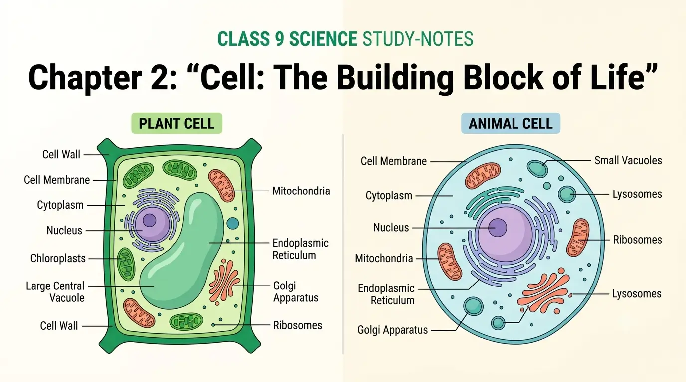
A plant cell has three extra structures that an animal cell does not have:
- Cell wall – an extra, rigid outer layer made of cellulose, outside the cell membrane. It gives shape and support.
- Plastids – such as chloroplasts, which contain chlorophyll for photosynthesis. This is why plants can make their own food.
- A large central vacuole – a big vacuole filled with cell sap. It stores water and helps keep the cell firm. In a mature plant cell it takes up a large part of the cell’s volume.
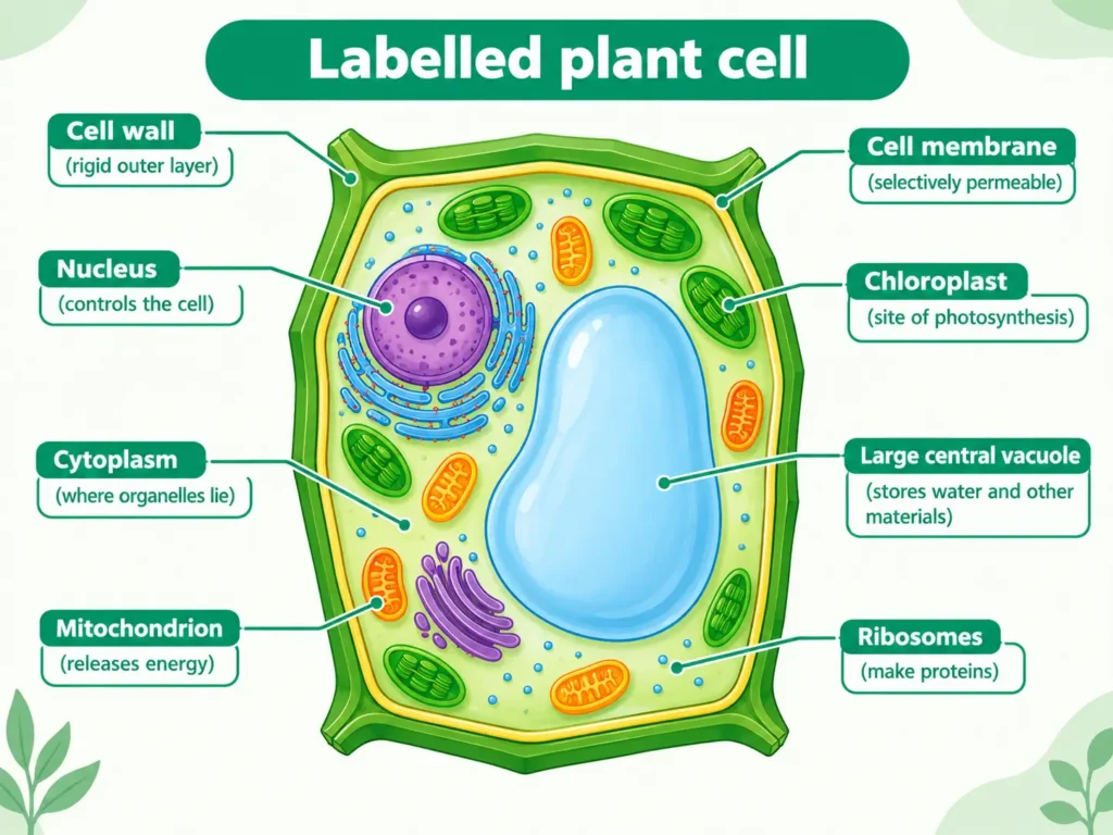
Animal cells have small, temporary vacuoles. They do not have a cell wall or plastids.
Beyond the main text (for general knowledge): Animal cells have a pair of structures called centrioles that help in cell division. Most higher plant cells do not have them. This detail is not needed for Class 9 answers, so do not write it unless a question asks.
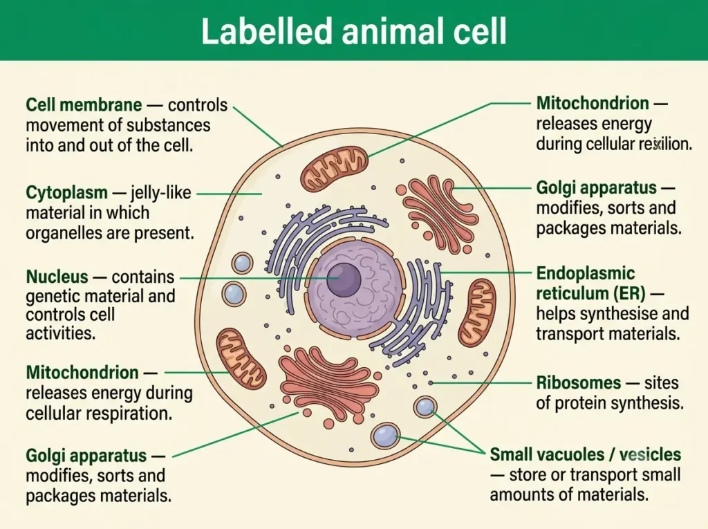
Common mistake: Saying the Golgi apparatus, ER or mitochondria are found only in animal cells. They are found in both plant and animal cells.
Exam point: Learn this table. Examiners often ask for two or three differences.
| Plant cell | Animal cell |
|---|---|
| Has a rigid cell wall outside the cell membrane | No cell wall; only the cell membrane |
| Has plastids such as chloroplasts | No plastids |
| Has a large central vacuole | Vacuoles are small and temporary |
| Usually box-like or regular in shape | Usually irregular or rounded in shape |
| Makes its own food by photosynthesis | Cannot make its own food |
Prokaryotic and Eukaryotic Cells
Scientists also classify cells by a second method: how well organised the nucleus is. The word prokaryotic comes from pro = primitive and karyon = nucleus. The word eukaryotic comes from eu = true and karyon = nucleus.
1. Prokaryotic cell – a cell that has no well-defined nucleus and no membrane-bound organelles.
- Typical size is about 1 to 10 µm.
- The DNA is a single, circular molecule lying directly in the cytoplasm in a region called the nucleoid. No membrane surrounds it.
- Membrane-bound organelles such as mitochondria, ER, Golgi apparatus and chloroplasts are absent. Most cell activities take place in the cytoplasm.
- Examples: bacteria and cyanobacteria (blue-green algae).
2. Eukaryotic cell – a cell that has a well-defined nucleus surrounded by a nuclear membrane, and membrane-bound organelles.
- Typical size is larger, about 10 to 100 µm.
- Examples: plant cells, animal cells, and the cells of fungi.
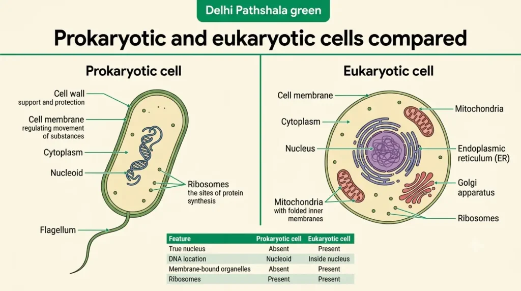
| Prokaryotic cell | Eukaryotic cell |
|---|---|
| Size about 1–10 µm | Size about 10–100 µm |
| No true nucleus; DNA lies in the nucleoid | True nucleus with a nuclear membrane |
| DNA is a single circular molecule | DNA is organised into several chromosomes with proteins |
| Membrane-bound organelles absent | Membrane-bound organelles present |
| Usually unicellular | Unicellular or multicellular |
| Example: bacteria | Example: plant and animal cells |
Exam point: Almost all prokaryotes are unicellular, while eukaryotes can be unicellular (Amoeba) or multicellular (humans). Write “usually unicellular” for prokaryotes, because some, like certain cyanobacteria, grow as chains or colonies.
Common mistake: Writing that “all cells have a nucleus”. Bacteria have only a nucleoid, and mature human red blood cells lose their nucleus to make room for haemoglobin.
Cell Organelles
A eukaryotic cell works like a tiny living factory. Different organelles carry out different jobs at the same time, and the whole cell stays alive because they work together. Let us go through each organelle one by one.
Nucleus
Definition: The nucleus is the organelle that contains the cell’s genetic material (DNA) and controls its activities. We often call it the brain of the cell.
Discovery: Robert Brown described the nucleus in 1831. It is a large, round body usually lying near the centre of the cell.
Structure (important for the exam)
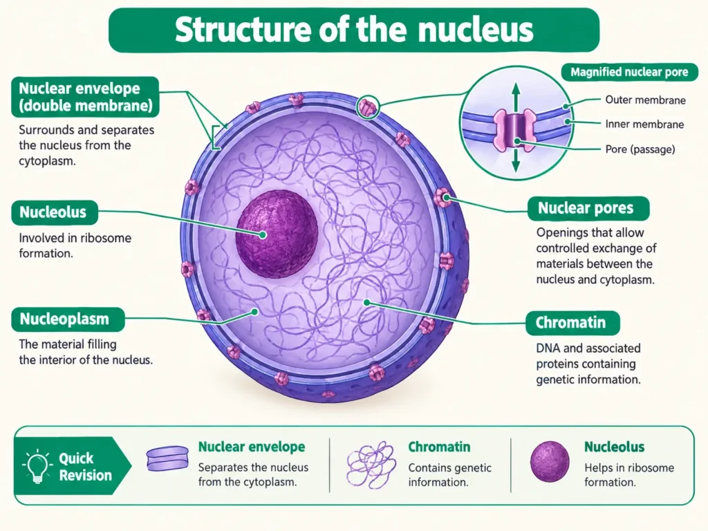
- It is surrounded by a double-layered nuclear membrane with small nuclear pores. The pores allow materials to pass between the nucleus and the cytoplasm.
- Inside is a fluid called nucleoplasm.
- A dark, dense, round body called the nucleolus is present. Ribosomal subunits are made here. They leave for the cytoplasm and join to form ribosomes.
- Chromatin is an entangled mass of thread-like structures made of DNA and proteins. It is seen in a cell that is not dividing. When the cell is about to divide, chromatin condenses into rod-shaped chromosomes.
- A functional segment of DNA is called a gene.
Functions you must remember
- It controls all cellular activities such as growth and division.
- It stores hereditary information as DNA, which passes from parents to offspring.
- It plays the main role in cell division.
Chain to memorise: cell → nucleus → chromosome → DNA → gene.
Exam point: Chromosomes carry the information for inheritance in the form of DNA.
Mitochondria
Have you ever wondered where a cell gets the energy to do its work? The answer is: from mitochondria. (The singular is mitochondrion; “mitochondria” is the plural.)
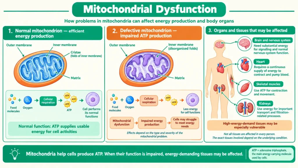
Definition: Mitochondria are the organelles in which food (such as glucose) is broken down in cellular respiration to release energy. They are called the powerhouse of the cell.
Structure
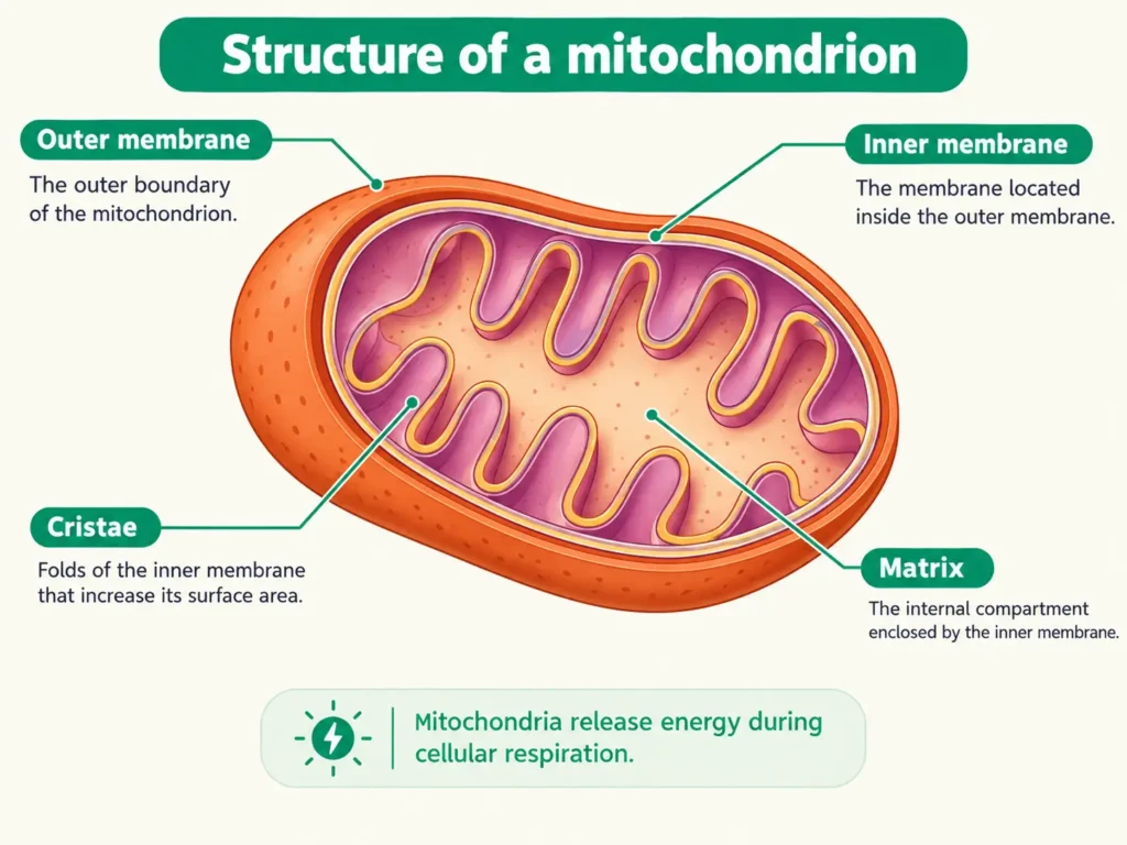
- They are covered by a double membrane. The outer membrane is smooth and porous. The inner membrane is folded into finger-like projections called cristae, which increase the surface area for chemical reactions.
- The inner space is filled with a fluid called the matrix, which contains its own DNA and ribosomes.
Functions
- The energy released is stored as ATP (Adenosine Triphosphate), called the energy currency of the cell.
- ATP energy is used for making new chemicals, movement and other work in the cell.
- Because mitochondria have their own DNA and ribosomes, they can make some of their own proteins. Strange, right? This also suggests that mitochondria and bacteria share a distant history.
Notice this carefully: cells that need a lot of energy, such as muscle cells, have many mitochondria.
Exam point: Mitochondria and plastids are the two organelles, other than the nucleus, that have their own DNA. A quick memory device: “M + P have their own DNA.”
Chloroplast
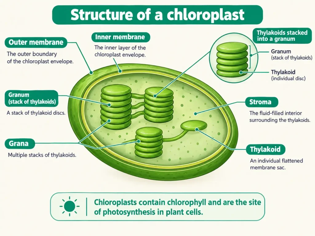
Definition: The chloroplast is a green plastid containing the pigment chlorophyll; it is the site of photosynthesis. If mitochondria are the powerhouse, the chloroplast is the kitchen of the cell.
Only plants (and algae) can make their own food, because only they have chloroplasts.
Structure
- Like mitochondria, chloroplasts have a double membrane.
- Inside is a semi-fluid substance called the stroma.
- In the stroma lie disc-shaped, coin-like structures called thylakoids, which contain chlorophyll. A stack of thylakoids is called a granum (plural: grana).
- Chloroplasts also have their own DNA and ribosomes.
Functions
- The main function is photosynthesis, in which a plant makes food (sugars) from carbon dioxide and water in the presence of sunlight.
- The green colour of leaves is due to chlorophyll in chloroplasts.
Other plastids (plastids are organelles for making and storing food in plant cells)
| Plastid | Colour | Function / example |
|---|---|---|
| Chloroplast | Green | Photosynthesis; found in leaves |
| Chromoplast | Yellow, orange or red | Gives colour to flowers and fruits, which attracts pollinators |
| Leucoplast | Colourless | Stores starch, oils or proteins; e.g., in potato |
Exam point: Chloroplasts are found in plant cells and in algae; they are not present in animal cells. Remember that bacteria such as cyanobacteria (blue-green algae) do photosynthesis but have no chloroplasts, because they are prokaryotes.
Mitochondria vs chloroplast
| Feature | Mitochondria | Chloroplast |
|---|---|---|
| Membranes | Double; inner membrane folded into cristae | Double; disc-shaped thylakoids in stroma |
| Own DNA and ribosomes | Yes | Yes |
| Function | Releases energy as ATP (respiration) | Makes sugars (photosynthesis) |
| Found in | Almost all eukaryotic cells | Photosynthetic plant cells |
Endoplasmic Reticulum
Look at the cytoplasm under a powerful (electron) microscope and you see a big network of tubes and sheets. That network is the Endoplasmic Reticulum (ER). Endoplasmic means inside the cytoplasm and reticulum means network.
Structure
- It is a large network of membrane-bound tubes and sheets spread through the cytoplasm.
- It is continuous with the outer membrane of the nuclear envelope.
- It has two types:
- Rough ER (RER) – ribosomes are attached to its surface, so it looks rough.
- Smooth ER (SER) – no ribosomes are attached, so it looks smooth.
Functions you must learn
- RER takes part in making and secreting proteins (for example, in the cells of the pancreas that secrete enzymes).
- SER helps in making and storing fats (lipids) and hormones.
- The ER acts as a transport system, carrying materials from one part of the cell to another.
- In liver cells, SER helps in detoxifying poisons and drugs.
Think of the ER like the roads inside a city. The RER is a road that carries protein “trucks”.
Vacuoles
Vacuoles are like the storage rooms of the cell. They are membrane-bound sacs filled with liquid.
Structure
- The membrane of the vacuole is called the tonoplast.
- The liquid inside is the cell sap, which contains water, sugars, minerals, amino acids and waste substances.
Plant cell versus animal cell (very important)
- A mature plant cell usually has one large central vacuole. It can occupy a very large part of the cell’s volume (more than half in many cells).
- Animal cells have small, temporary vacuoles, often many in number.
Functions
- Storage of water, food, minerals, pigments and waste products.
- In plant cells, the vacuole maintains turgor pressure, the pressure that keeps leaves and stems firm. When a plant loses water, the vacuole shrinks and the plant wilts.
- Pigments in vacuoles give colour to some flowers and fruits.
- In unicellular organisms like Amoeba, vacuoles help in taking in food (food vacuole) and removing excess water (contractile vacuole).
Exam point: Why are vacuoles large in plant cells? To store water and other materials and to give the cell firmness and support.
Plasma Membrane
Every cell has an outer boundary. In animal cells it is the outermost layer; in plant cells it lies just inside the cell wall. This boundary is called the plasma membrane or cell membrane.
Definition: The plasma membrane is a thin, flexible, selectively permeable layer that surrounds the cell, defines it and protects its contents.
Structure
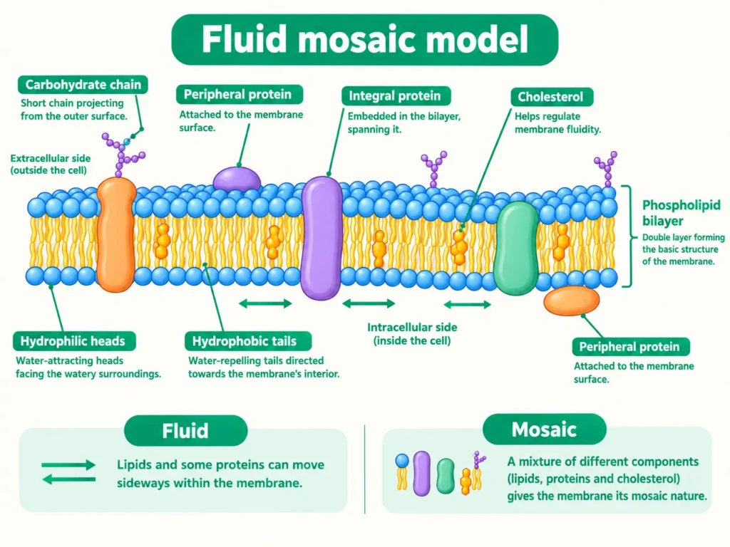
- It is made mainly of lipids and proteins.
- It is selectively permeable, which means it allows some substances to pass through while blocking others. (You may also see the term semi-permeable used for this idea.)
- The Fluid Mosaic Model explains its structure. The lipids form a double layer (lipid bilayer), and proteins are embedded in it. The molecules can move sideways, so the membrane is fluid, and they are arranged like tiles in a mosaic, so it is a mosaic.
Functions (very important for 2-mark questions)
- It gives shape to the cell and protects it.
- It controls what enters and leaves the cell, keeping useful substances in and harmful ones out.
- It helps in diffusion and osmosis. For example, oxygen enters the cell through this membrane.
- Because it is flexible, some cells (such as Amoeba) can engulf food by a process called endocytosis.
Think of a gatekeeper at your school gate. He checks each person and lets in only those who should enter. The plasma membrane does the same job.
Cell Wall
You already know that plant cells have something extra outside the plasma membrane. That is the cell wall.
Definition: The cell wall is a rigid, permeable outer layer present in plant cells, fungi and bacteria. It is absent in animal cells.
Structure
- It is mainly made of cellulose, a carbohydrate formed of many glucose units linked together. It gives the wall strength.
- It is non-living and permeable: water and dissolved minerals pass through it freely. The plasma membrane just inside it is the structure that is selectively permeable.
- Plant cells are connected to each other by fine cytoplasmic channels called plasmodesmata, which pass through the walls. (This is an extra detail beyond the main textbook text.)
Functions
- It gives rigidity, shape and strength to the cell. That is why plants can stand tall without bones.
- It protects the cell from mechanical injury.
- It prevents the cell from bursting when water enters, so plant cells can take in a lot of water without breaking, while animal cells would burst.
- It helps to hold the plant’s structure firm; leaves and flowers stay upright.
A useful observation: Put a plant cell (such as onion or Rhoeo) in concentrated sugar solution. The cell contents shrink and the membrane pulls away from the wall, but the outer outline stays the same because the wall is rigid. An animal cell (such as a cheek cell) has no wall, so it shrinks much more. This flexibility lets animal cells change shape easily.
Plasma membrane vs cell wall (remember this table for the exam)
| Plasma membrane | Cell wall |
|---|---|
| Present in all cells | Present only in plant cells, fungi and bacteria |
| Made of lipids and proteins; living | Mainly cellulose (in plants); non-living |
| Selectively permeable | Permeable |
| Thin and flexible | Thick and rigid |
| Lies inside the cell wall (in plants) | Lies outside the plasma membrane |
Ribosomes
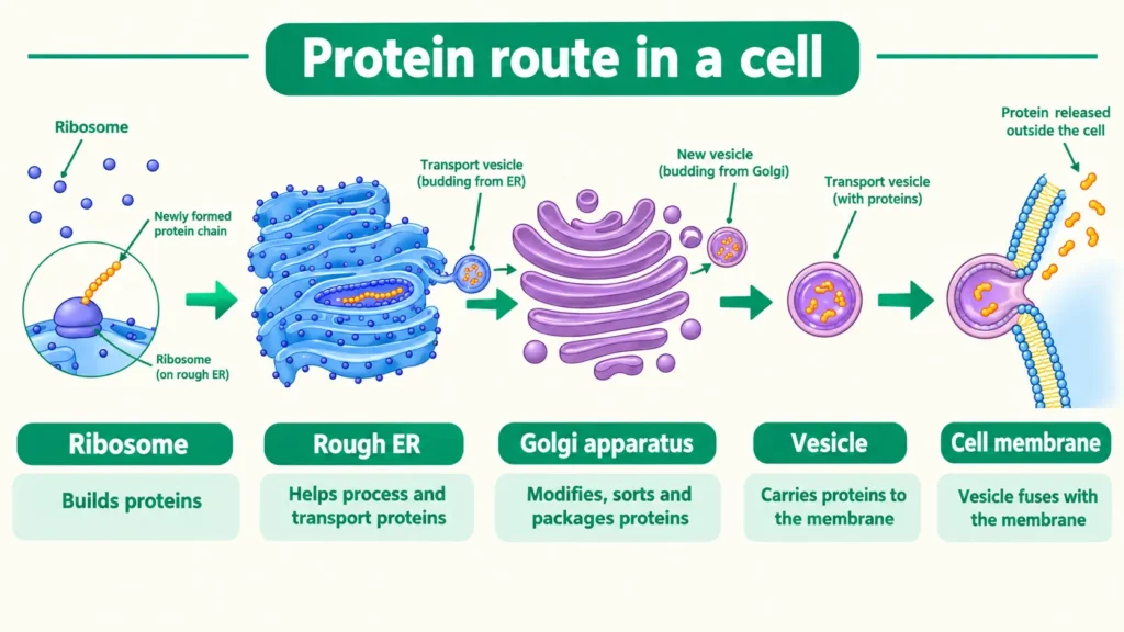
Ribosomes are tiny structures found either free in the cytoplasm or attached to the rough ER. They are the sites of protein synthesis, which is why they are called the protein factories of the cell. They are present in both prokaryotic and eukaryotic cells.
Golgi Apparatus
The Golgi apparatus is a stack of flattened, sac-like structures linked to the ER and to the cell membrane. It modifies, sorts and packages proteins and lipids into small sacs called vesicles, which carry them to different places inside the cell or out of it. It also helps in forming lysosomes. It is often called the packaging and dispatch centre (or post office) of the cell. It was first described by the Italian scientist Camillo Golgi in 1898.
The protein route to remember: Ribosome (makes the protein) → RER (folds and processes it) → Golgi apparatus (packages it into vesicles) → cell membrane (the protein is exported).
Lysosomes
Lysosomes are small sacs, bound by a single membrane, filled with powerful digestive enzymes. They break down unwanted proteins, carbohydrates, fats and worn-out parts of the cell, and the products are reused. They are called the clean-up system of the cell. When a cell is damaged or old, lysosomes can burst and digest the cell itself. This is why they are nicknamed the “suicide bags” of the cell.
Cytoplasm and Other Parts
The cytoplasm is the semi-fluid, jelly-like substance between the plasma membrane and the nucleus. All organelles lie in it. Many cells also contain a network of fine fibres called the cytoskeleton, which gives support, helps in keeping the shape, and helps in movement and transport inside the cell. Stored materials such as starch or crystals are called cell inclusions. (These two terms are enrichment points; learn them only if your teacher asks.)
Organelles at a glance
| Organelle | Nickname | Key function | Plant / animal |
|---|---|---|---|
| Nucleus | Brain of the cell | Controls activities; stores DNA | Both |
| Mitochondria | Powerhouse | Releases energy as ATP | Both |
| Chloroplast | Kitchen | Photosynthesis | Plant only |
| Ribosome | Protein factory | Protein synthesis | Both |
| ER | Transport network | Protein/lipid synthesis; transport | Both |
| Golgi apparatus | Packaging unit | Modifies and packs proteins and lipids | Both |
| Lysosome | Clean-up / suicide bag | Digests waste and old parts | Both |
| Vacuole | Storage room | Stores water and materials; gives firmness | Large in plants, small in animals |
| Plasma membrane | Gatekeeper | Controls entry and exit | Both |
| Cell wall | Protective armour | Shape, strength, protection | Plants, fungi, bacteria |
Permeability of Cell Membranes
Diffusion and Osmosis
You have learnt that the plasma membrane is selectively permeable. It does not allow everything to pass; it checks what goes in and out. This property is called permeability, and because of it two very important processes take place: diffusion and osmosis.
1. Diffusion
Have you noticed how the smell of perfume spreads from one corner of the room to the whole room? That is diffusion.
Definition: Diffusion is the net movement of particles from a region of higher concentration to a region of lower concentration.
- It occurs in gases and liquids (and for dissolved substances).
- It does not need a membrane. It can happen in open air too.
- Example: Oxygen moves into the cell, where its concentration is lower, and carbon dioxide moves out of the cell in the same way. Both cross the plasma membrane by diffusion.
2. Osmosis
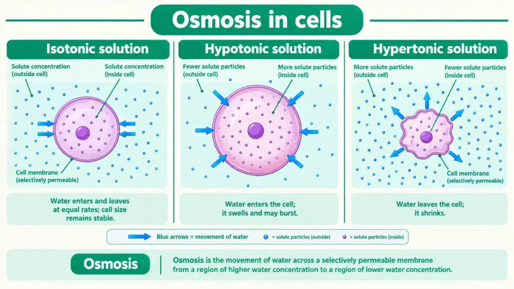
Definition: Osmosis is the diffusion of water across a selectively permeable membrane. Water moves from a more dilute solution (more water) to a more concentrated solution (less water) until the concentrations become equal.
Notice this carefully: osmosis always needs a selectively permeable membrane. In plants, water from the soil enters the root cells by osmosis.
Isotonic, hypotonic and hypertonic solutions
| Solution (outside the cell) | Solute concentration compared with the cell | Water movement | What happens to the cell |
|---|---|---|---|
| Hypotonic (e.g., plain water) | Lower outside | Water enters the cell | Cell swells |
| Hypertonic (e.g., strong salt or sugar solution) | Higher outside | Water leaves the cell | Cell shrinks |
| Isotonic | Same as inside | No net movement | Cell stays the same size |
The NCERT activity: potato pieces and osmosis
Take two similar pieces of potato. Weigh them. Keep one in plain water and the other in a 20% salt (or sugar) solution. After some time, weigh them again.
- The piece in plain water swells and gains weight, because water enters its cells by osmosis (plain water is hypotonic to the cells).
- The piece in the concentrated solution shrinks and loses weight, because water leaves its cells by osmosis (the solution is hypertonic).
- The membrane lets water pass but not the large salt or sugar molecules. This proves osmosis.
The same idea is often shown with dried raisins (they swell in plain water and shrink in concentrated sugar solution), and your school exam may use either example.
Real-life link: Hospitals give patients isotonic saline drips. The saline has the same solute concentration as blood, so red blood cells neither swell nor shrink.
Common mistake: Writing that “salt enters the cell” in the potato experiment. Only water moves; the salt molecules do not cross the membrane.
Diffusion vs osmosis
| Diffusion | Osmosis |
|---|---|
| Movement of particles of any substance (gas, liquid, dissolved) | Movement of water only |
| No membrane needed | Needs a selectively permeable membrane |
| From higher to lower concentration of the substance | From higher to lower water concentration (dilute to concentrated solution) |
Why is it important? Cells get water and nutrients and remove waste because of diffusion and osmosis. Without osmosis, roots could not absorb water from the soil.
Cellular Division and Cancer
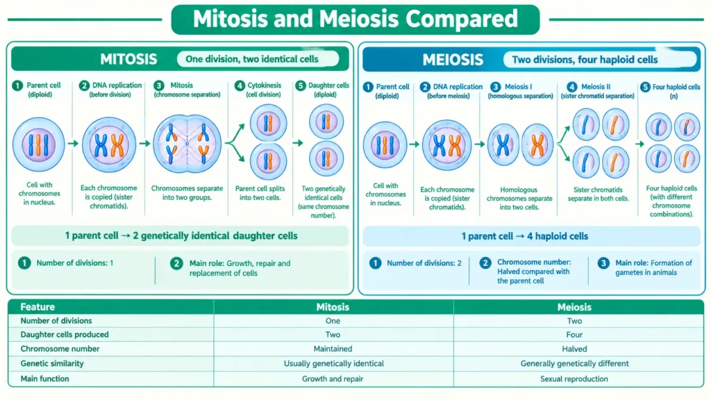
Mitosis and Meiosis
New cells arise from old cells. This is how you grew from a small baby into a Class 9 student. A parent cell divides to form new cells in a process called cell division, which is needed for growth, repair of wounds and reproduction. Remember Virchow’s idea: all cells arise from pre-existing cells.
In eukaryotic cells, division takes place in a controlled and orderly way through a series of events known as the cell cycle. In Class 9 you study two kinds of cell division.
1. Mitosis (also called equational division)
- It takes place in body (somatic) cells such as skin, blood and bone cells.
- One parent cell divides to form two identical daughter cells, each with the same number of chromosomes as the parent.
- It is used for growth and repair; when you get a cut, new skin cells are made by mitosis. It is also the basis of asexual reproduction.
- For general knowledge: The stages of mitosis are prophase, metaphase, anaphase and telophase. You are not required to describe them in detail in Class 9.
2. Meiosis (also called reductional division)
- It takes place in reproductive cells: in the testes and ovaries, to form sperms and eggs (gametes).
- One parent cell divides twice to form four daughter cells, each with half the number of chromosomes of the parent. A human body cell has 46 chromosomes, but a sperm or an egg has 23.
- When a sperm (23) and an egg (23) fuse, the baby again has 46.
- Meiosis is essential for sexual reproduction, and it also brings variation.
Mitosis vs meiosis (examiners often ask this for 3 marks)
| Mitosis | Meiosis |
|---|---|
| Occurs in body cells | Occurs in reproductive cells |
| Forms 2 daughter cells | Forms 4 daughter cells |
| Chromosome number remains the same | Chromosome number becomes half |
| Daughter cells are identical to the parent | Daughter cells differ from the parent, bringing variation |
| Helps in growth and repair | Helps in forming gametes |
| Equational division | Reductional division |
Common mistake: Writing that meiosis forms two cells. It forms four.
Exam point: Mitosis for growth and repair; meiosis for reproduction.
Cancer
Normally, cell division is carefully controlled. Cells divide only when the body needs them to, and many normal cells stop dividing when they touch their neighbours (called contact inhibition). Old or damaged cells are removed by a controlled process of programmed cell death.
Sometimes this control is lost, and cells divide again and again without stopping. This uncontrolled division of cells is called cancer, and the mass of cells it forms is called a tumour. For Class 9, you only need this general understanding; errors in the division of body cells (mitosis) can lead to tumours, whereas errors in meiosis can cause genetic disorders in offspring.
Recent Advancement in Cell Biology
Case Study and Indian Contribution
In Class 9 you learn the basic structure of the cell. But cell biology is not a closed chapter. Scientists continue to make new discoveries, and today we can study cells with very powerful microscopes and even read and edit the instructions (DNA) inside them.
For this chapter, the syllabus asks you to understand three ideas: how problems in a cell organelle can cause disease (the Leigh syndrome case study), that Indian scientists have made major contributions (for example, Prof. Arun Kumar Sharma), and that science keeps raising new questions (for example, can we make an artificial cell?).
Notice this carefully: The Delhi Directorate of Education annual syllabus for Class 9 Science (2026-27), which follows the same textbook, names Leigh syndrome and Prof. Arun Kumar Sharma as examples for this chapter, so these are not “extra” topics. They are usually asked as short answers or as part of a case-based question.
Leigh Syndrome and Mitochondrial Dysfunction
You have learnt that mitochondria are the powerhouse of the cell because they make energy in the form of ATP. What happens if the powerhouse does not work properly? The cell does not get enough energy. This is what happens in mitochondrial dysfunction.
Leigh syndrome is a rare, inherited disorder caused by faulty mitochondria.
- It usually shows up in infancy or early childhood, although later onset has been reported in some older children and adults.
- Doctors have found that it can be caused by changes (mutations) in mitochondrial DNA or in nuclear DNA, which affect the proteins that mitochondria need to make energy.
- The nervous system is mainly affected, because nerve cells need a lot of energy. Children may lose abilities they had gained, have difficulty in movement, feeding or swallowing, and may show symptoms such as vomiting or irritability.
- Its older medical name is subacute necrotizing encephalomyelopathy. You do not need to remember this long name for Class 9.
- According to the medical sources checked for these notes, there is no cure that changes the course of the disease; doctors mainly manage the symptoms.
Why is this example important? It shows the link between a single cell organelle and human health. If the mitochondria fail, cells run short of ATP, and the organs that need the most energy suffer first. This is why organelles are called functional parts of the cell.
Exam point: Name a disease caused by mitochondrial dysfunction. Leigh syndrome.
Contribution of Prof. Arun Kumar Sharma
For an Indian contribution to cell biology, the textbook syllabus gives the example of Prof. Arun Kumar Sharma, who is considered by many to be the father of Indian cytology (cytology means the study of cells).
- He was born on 31 December 1924 in Kolkata and worked mainly at the University of Calcutta, where he headed the Department of Botany and set up the Centre of Advanced Study in Cell and Chromosome Research.
- His main work was on chromosomes: their structure, their chemistry and the methods of studying them in plants.
- With his wife and fellow scientist Archana Sharma, he developed new methods of treating and staining cells so that chromosomes could be seen clearly. Together they wrote the book Chromosome Techniques: Theory and Practice (1965), used by students and researchers worldwide.
- He proposed a new concept of speciation in plants that reproduce vegetatively (without seeds).
- He received the Shanti Swarup Bhatnagar Prize (1967) and the Padma Bhushan (1983).
Exam point: If asked for an Indian scientist in cell biology, you can write: Prof. Arun Kumar Sharma studied chromosomes and developed techniques for studying them, particularly in plants. He is considered the father of Indian cytology.
His work shows that careful observation and good techniques, not only costly instruments, can lead to important discoveries.
Can We Make an Artificial Cell?
The textbook ends its discussion by encouraging you to ask questions such as: Can we create an artificial cell that behaves exactly like a natural living cell? A step in this direction came in 2010, when a team led by J. Craig Venter put chemically made DNA of a bacterium into another bacterial cell from which the original DNA had been removed. The cell grew and divided under the instructions of the new DNA, showing that DNA controls the structure and activities of a cell. Scientists still cannot build a living cell from non-living parts, and if one were made, many ethical questions would arise. Think about these questions yourself; they can appear as reasoning questions.
Practical: Preparing Stained Temporary Mounts
Preparing Stained Temporary Mounts of Onion Peel and Human Cheek Cells
This is an important practical for your school exams. You make two temporary mounts, one of onion peel and one of human cheek cells. Steps are often asked for 2 marks, so learn them in order.
Aim: To prepare stained temporary mounts of onion peel and human cheek cells and to study their structure.
Materials required: Onion, forceps, blade, watch glass, glass slide, coverslip, needle, brush, blotting paper, microscope, safranin stain, methylene blue stain, glycerine, and water.
A. Mount of onion peel
- Take an onion and break off a thick fleshy leaf. With the forceps, peel off a thin, transparent skin from the inner side. This is the epidermal peel.
- Put the peel in a watch glass with water so it does not dry up.
- Add 1–2 drops of safranin stain. Leave it for about a minute. It stains the cell wall and nucleus red/pink.
- With a brush, transfer the peel to a clean glass slide. Add a drop of glycerine.
- Lower the coverslip gently with the help of a needle to avoid air bubbles. Remove extra glycerine with blotting paper.
- Observe under the microscope, first on low power and then on high power.
B. Mount of human cheek cells
- With a clean, blunt tooth-pick or ice-cream stick, gently scrape the inside of your cheek. Use a fresh stick, use it only once and dispose of it properly.
- Spread the material on a clean glass slide in a drop of water.
- Add 1–2 drops of methylene blue stain, which stains the nucleus blue.
- Put on the coverslip carefully (avoiding air bubbles), and observe under the microscope.
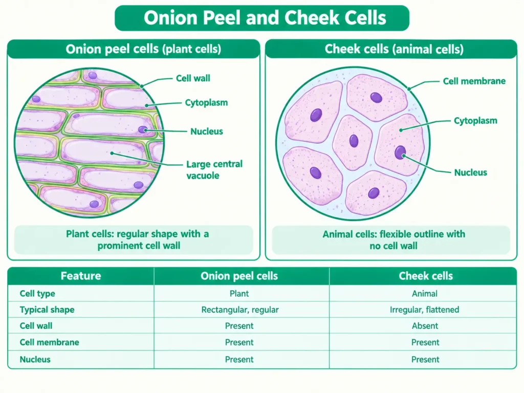
Observations: Onion cells are box-shaped (rectangular) and regularly arranged, and each has a clear cell wall, a nucleus and a large vacuole. Cheek cells are irregular or rounded, with no cell wall. Both have a nucleus and cytoplasm.
Conclusion: Plant cells have a cell wall and animal cells do not; both have a nucleus, cytoplasm and a cell membrane.
Precautions: The peel should be thin and small. Do not let the mount dry. Avoid air bubbles under the coverslip. Handle the blade and needle carefully. Wash your hands after the practical.
(Check with your teacher for the exact experiment number in your school’s practical list; it may differ from older notes.)
Conclusion
The cell is the unit in which life is built and run. From Hooke’s dead cork cells to modern electron microscopes, our picture of the cell has become clearer and clearer. Remember the main thread: cells can be prokaryotic or eukaryotic; the plasma membrane controls what enters and leaves; organelles share the work like departments in a factory; osmosis and diffusion keep the cell supplied; and cells make more cells by mitosis and meiosis, under control, unless something goes wrong, as in cancer. Cases like Leigh syndrome remind us that the health of a whole person depends on tiny organelles working properly.
Quick Summary
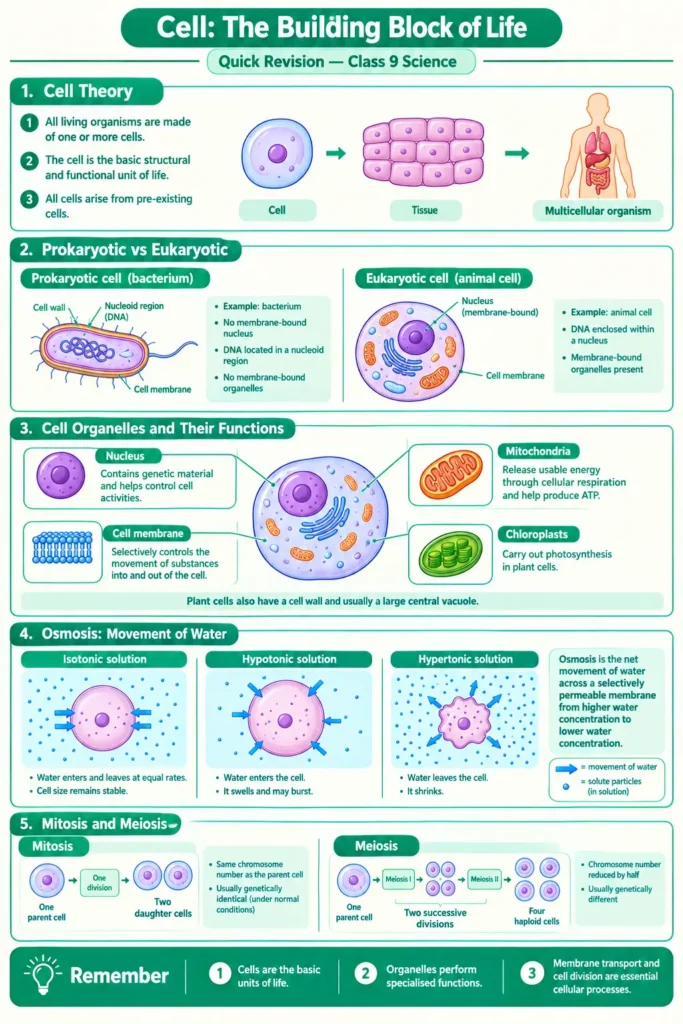
- Cell theory: All living things are made of cells; the cell is the basic structural and functional unit of life; all cells come from pre-existing cells (Hooke 1665, Schleiden–Schwann 1838–39, Virchow 1855).
- Cell types: Prokaryotic cells have a nucleoid and no membrane-bound organelles; eukaryotic cells have a true nucleus and organelles. Plant cells have a cell wall, plastids and a large vacuole in addition.
- Organelles: Nucleus is the control centre; mitochondria make ATP; chloroplasts make food; ribosomes make proteins; ER transports; Golgi packages; lysosomes digest; vacuoles store; the plasma membrane is selectively permeable; the cell wall is rigid and permeable.
- Movement and division: Diffusion is movement from higher to lower concentration; osmosis is the diffusion of water across a selectively permeable membrane; mitosis gives two identical cells for growth and repair; meiosis gives four cells with half the chromosomes for reproduction; uncontrolled division is cancer.
NCERT-Style Questions with Answer Points
Note: The questions below are the well-known classic questions on the cell. They are written in “NCERT-style” for practice. They are not copied from the 16 Revise, Reflect, Refine questions at the end of the new 2026-27 chapter, whose wording I could not check. For the exact questions, use your textbook.
Q1. Who discovered cells and how? Robert Hooke discovered cells in 1665. He observed a thin slice of cork under a microscope he had designed himself and saw small box-like compartments, which he named cellulae.
Q2. Why is the cell called the structural and functional unit of life? Structural unit: all living organisms are made of cells, like bricks make a building. Functional unit: all life functions such as respiration, digestion and excretion take place inside cells.
Q3. How do substances like CO₂ and water move in and out of the cell? CO₂ moves by diffusion, from a higher concentration to a lower concentration, across the plasma membrane. Water moves by osmosis, across the selectively permeable plasma membrane, from the more dilute side to the more concentrated side.
Q4. Why is the plasma membrane called a selectively permeable membrane? Because it allows only some substances to enter and leave the cell, keeping useful substances in and harmful ones out.
Q5. Name the two organelles that contain their own genetic material. Mitochondria and plastids (such as chloroplasts). They contain their own DNA and ribosomes, so they can make some of their own proteins.
Q6. If the organisation of a cell is destroyed by some physical or chemical influence, what will happen? The cell will not be able to carry out its basic functions such as respiration, energy release and protein synthesis, because the organelles work together. If the coordination is lost, the cell will die.
Q7. Why are lysosomes known as suicide bags? They contain powerful digestive enzymes. When the cell is damaged or old, lysosomes can burst and digest the cell itself.
Q8. Where are proteins synthesised inside the cell? In the ribosomes, which are free in the cytoplasm or attached to the rough ER.
Q9. What is the function of the Golgi apparatus? It modifies, sorts and packages proteins and lipids made in the ER into vesicles and sends them to different parts of the cell or out of it. It also helps in forming lysosomes.
Frequently Asked Questions
Why do plant cells not shrink as much as animal cells in concentrated sugar solution?
The rigid cell wall keeps the outer shape of a plant cell. Its contents shrink and the membrane pulls away from the wall, but the outline stays. A cheek cell has no wall, so it shrinks visibly.
What is the difference between chromatin and chromosomes?
They are the same material in two forms. In a cell that is not dividing, DNA and proteins appear as thin, entangled chromatin. When the cell is about to divide, the chromatin condenses into rod-shaped chromosomes.
Do bacteria have a nucleus?
No. Bacteria are prokaryotes. Their DNA is a circular molecule lying in the nucleoid, and no membrane surrounds it.
Is osmosis the same as diffusion?
Osmosis is a special case of diffusion. Diffusion can involve any particles and needs no membrane; osmosis is only the movement of water across a selectively permeable membrane.
Why can mitochondria and chloroplasts make some of their own proteins?
Both have their own DNA and ribosomes. This also suggests that they share a distant evolutionary history with bacteria.
Does a mature red blood cell have a nucleus?
No. Human red blood cells lose their nucleus as they mature, which leaves room for haemoglobin. They cannot divide and live for about 120 days.
Which structures are found only in plant cells?
Chloroplasts and other plastids, and the large central vacuole, are typical of plant cells. The cell wall is present in plants, fungi and bacteria, but not in animal cells. The nucleus, mitochondria, ER, Golgi apparatus and ribosomes are found in both plant and animal cells.
Is Leigh syndrome part of the Class 9 syllabus?
Yes, as a case study. The 2026-27 syllabus names it as an example of the link between mitochondrial dysfunction and human health. You only need the idea, not the medical details.
External References
External References — Class 9 Science, Chapter 2
Subscribe to keep reading
You've used your free notes for now. Get Delhi Pathshala Premium for unlimited access.
Subscribe — ₹29/30 days